Science Photo Library

Cancer learning ‘hub’
Pharmacists are playing an increasingly important role in supporting patients with cancer, working within multidisciplinary teams and improving outcomes. However, in a rapidly evolving field with numbers of new cancer medicines is increasing and the potential for adverse effects, it is now more important than ever for pharmacists to have a solid understanding of the principles of cancer biology, its diagnosis and approaches to treatment and prevention. This new collection of cancer content, brought to you in partnership with BeOne Medicines, provides access to educational resources that support professional development for improved patientEpidemiology
Data from 2016 show that lung cancer is the most common cause of cancer death in the UK, accounting for 21% of all cancer deaths, and UK crude mortality rates show 62 lung cancer deaths per 100,000 males and 50 per 100,000 females per year[1] . Lung cancer survival in the UK has improved little over the past 40 years and, of the 20 most common cancers in the UK, the 10-year survival ranks second lowest, with only 5% of people with lung cancer in England and Wales surviving 10 years following diagnosis (data from 2010–2011). Survival rates for lung cancer are strongly related to stage of disease at diagnosis, with one-year survival for lung cancer highest in patients diagnosed at stage 1 (83%) compared with the lowest in stage 4 (17%)[1] . However, these statistics combine the two major defined histological groups of lung cancer: small-cell lung cancer (SCLC) and NSCLC. Survival rates differ between the two groups, and are dependent on stage at diagnosis. Histology now plays a much larger part in defining the subtype of lung cancer, which subsequently influences treatment choices and survival rates.Treatment algorithm
There is a variety of treatment modalities for lung cancer including surgery, radiotherapy and systemic anticancer treatment (SACT), but choice of treatment depends on stage of cancer at diagnosis, performance status and patient preference. These factors are discussed fully in the National Institute for Health and Care Excellence (NICE) guidance ‘Lung Cancer: Diagnosis and Management’ and the current NICE Lung Cancer Pathways[2] . However, these guidelines and pathways date from 2011. They are currently under review with an updated version expected in March 2019.Surgery
Patients with early-stage lung cancer have the option to undergo surgical resection, accounting for 20–25% of those in this group. There have been advances in surgical techniques, such as the widespread uptake of video-assisted thoracoscopic surgery (VATS) in the 1990s, in place of conventional thoracotomy (open surgery). In VATS the surgeon inserts surgical instruments and a camera through small incisions in the thorax, whereas in conventional surgery, a much larger incision is made. Compared with thoracotomy, VATS has resulted in better preservation of pulmonary function[3] and overall surgical mortality of 0–2%[4],[5] . More recent advancements have focused on the possible benefits of single-port VATS and the introduction of robotic-assisted thoracoscopic surgery (RATS). Unfortunately, owing to the comorbidity often associated with smoking and the age of onset of lung cancer, 20–30% of patients diagnosed with early-stage lung cancer are considered unfit for surgery. The median survival for patients with untreated T1 tumours (tumours <3cm in size) is 13 months[6] and, as such, radiotherapy is warranted in this group of inoperable patients.Radiotherapy and chemotherapy
In patients with tumours that are inoperable owing to more advanced stages of disease, radiotherapy is used. Conventional radiotherapy involves fractionated radiation of 2.0–2.75Gy/day for a total biologically equivalent radiation dose of 60-70Gy, which equates to around six weeks of treatment. The current NICE guidance for radical radiotherapy recommends either the continuous hyperfractionated accelerated radiotherapy (CHART) regimen (36 fractions of 1.5 Gy, 3 times per day) or, if CHART is not available, conventionally fractionated radiotherapy (64–66Gy in 32–33 fractions over 6.5 weeks or 55Gy in 20 fractions over 4 weeks). However, there is variation both nationally and internationally on the final dosage and sequencing of the radiotherapy. Chemotherapy can be used prior to, or concurrently with, radiotherapy. Several trials, including the pivotal RTOG9410 trial[7] , show an advantage to concurrent chemoradiotherapy treatment (which is the international standard of care when achievable). Positive results for accelerated schedules such as the UK-based CHART technique[8] have been demonstrated and there is potential for benefit with dose escalation. However, this is tempered by potential adverse outcomes as shown in RTOG0617[9] , which was closed early owing to lack of benefit. Modern techniques, such as 4D radiotherapy planning combined with intensity modulated radiation therapy, are now utilised to reduce toxicity. However, the evidence is clear that in stage 1 lung cancer, conventional radiotherapy is inferior to surgery, with local recurrence found to be, at worst, up to 70%[6] . In attempts to improve these results, stereotatic ablative radiotherapy (SABR), a technique that combines stereotactic localisation with high-dose hypofractionation, has been used. In general, 3–5 fractions are delivered in a period of less than two weeks. This concentration results in much higher ablative doses of radiation and is thought to provide similar local control to surgical resection. In recent years, SABR has been considered the treatment of choice for medically inoperable patients with early-stage NSCLC[10] . Despite the advances in surgery and radiotherapy, use of these treatments, such as monotherapy, is generally restricted to early-stage disease; SACT is the mainstay of treatment for advanced stage 3b–4 NSCLC and is the preferred initial treatment for both limited and extensive SCLC. The main indications for SACT in the advanced lung cancer population is to improve survival, disease control and quality of life. The rest of this article focuses on the use of SACT.Emerging treatments
Histology to guide treatment choice
Initially, the classification for lung cancer was divided into large cells accounting for 90% of cases and small cells accounting for the remainder. These two groups behave quite differently in their growth rate, pattern of spread and response to available treatment. Later, further subtyping occurred within NSCLC into adenocarcinoma, the cells of which form glandular-type structures similar to the mucous-producing lung tissue, and squamous cell carcinoma, which resemble the flat cells lining the airways. Advances in immunohistochemistry and molecular and genetic techniques now allow identification of many other rarer cell types. In the UK lung cancer population, the most prevalent histology is adenocarcinoma (36%), followed by not pathologically confirmed (31%), squamous (22%), SCLC (10%) and other (11%)[11] . The percentage of patients whose lung cancer is not pathologically confirmed illustrates the morbidity of the lung cancer population where fitness to treat and, indirectly safety of biopsy, is a major determinant of outcome. In 2002, a landmark trial conducted by the European Oncology Cooperative Group (ECOG), that compared four first-line platinum-based chemotherapy doublets in patients with advanced NSCLC was published[12] . The trial showed no difference in overall survival between the regimens, which led a perception that there was no requirement to refine patient selection other than by ECOG performance status. This perception was challenged on the publication of a phase III trial comparing cisplatin-pemetrexed and cisplatin-gemcitabine in advanced NSCLC[13] . Sub-group analyses showed different activity between the adnenocarcinomas and squamous groups, with the adenocarcinomas having significantly longer overall survival in the pemetrexed arm. However, in the squamous patient group, overall survival was significantly longer in the gemcitabine arm. This led to the approval of pemetrexed for non-squamous cancers only and led to the development of histological subtyping in NSCLC.Molecular targets
Epidermal growth factor receptor
The first clinically relevant molecular target in NSCLC to be identified was the epidermal growth factor receptor (EGFR) mutation. Oral EGFR tyrosine kinase inhibitors (TKIs; e.g. gefitinib, erlotinib and afatanib) block the EGFR mutations in the tyrosine kinase domain (Figure 1). The most common EGFR mutations detected are L858R point mutation in exon 21 and exon 19 deletion[14] . The progression-free survival (PFS) of TKIs when compared with platinum-based chemotherapy in EGFR+ patients was significantly superior (9–13 months vs. 5–7 months)[15],[16],[17] combined with response rates of 75%. TKIs are now the first-line option for EGFR+ patients with non-squamous NSCLC, with routine testing for EGFR mutations undertaken prior to SACT being initiated. Gefitinib, erlotinib and afatanib are all available under NICE technology appraisals for the first-line treatment of patients with EGFR+ NSCLC[18],[19],[20] . However, prevalence of EGFR+ mutations limits the widespread use within UK practice, with only 10–15% of all adenocarcinomas in the Caucasian population being EGFR+ compared with up to 60% of Asian populations[21] . 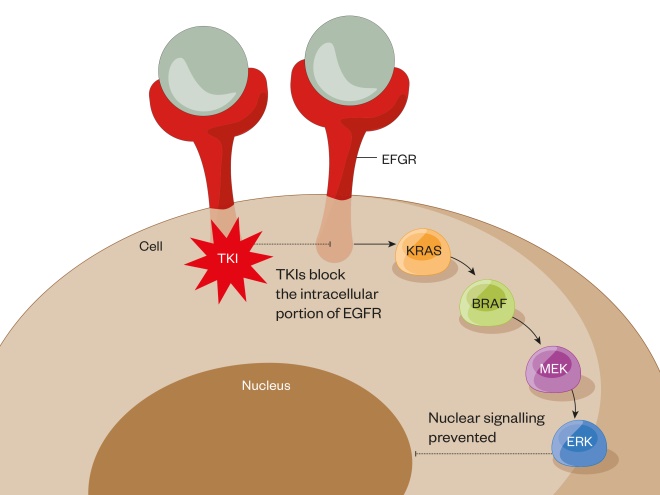
Figure 1: EGFR TKIs mechanism of action
EGFR = epidermal growth factor receptor; TKI = tyrosine kinase inhibitor
Oral epidermal growth factor receptor (EGFR) tyrosine kinase inhibitors (TKIs), such as gefitinib, erlotinib and afatanib, block the EGFR mutations in the tyrosine kinase domain.
Anaplastic lymphoma kinase
A second molecular target is known for NSCLC — ALK mutations. This mutation is rarer than EGFR mutations, contributing to around 1–4% of all NSCLC cases. Two oral ALK TKIs, crizotinib and ceritinib, are available under NICE technology appraisals[27],[28] , while alectinib may become available for more patients in the foreseeable future[29] (it is currently only available for NHS patients under the compassionate use scheme). Crizotinib showed significantly improved PFS in ALK+ patients, whether patients were pre-treated with platinum combinations (7.7 months vs. 3.0 months)[30] or in the first-line setting (10.9 months vs. 7.0 months)[31] . As with EGFR mutations, resistance mechanisms lead to the failure of ALK TKIs and second-generation ALK TKIs (e.g. ceritinib), which have been shown to be effective in those patients who are crizotinib resistant. Unlike EGFR mutations, however, there is no requirement to identify the resistance mechanism in practice[32] . Ceritinib has also been shown to have activity in crizotinib-naive patients[33] and both are now available for use in the first-line setting. There are no data available for a direct comparison between these two agents, but indirectly there is evidence for improved PFS for ceritinib versus crizotininb when compared with chemotherapy; however, it is not clear how the sequencing of these agents affects survival. A very recent head-to-head comparator study of alectinib versus crizotinib in the first-line setting demonstrated superior PFS for alectinib and lower overall toxicity rates, although the median PFS with alectinib was not reached[34] . The identification of EGFR and ALK mutations has led to the upfront testing for these mutations in advanced non-squamous NSCLC patients and drastically changed the treatment algorithm (Figure 2) in these patients. The effectiveness of these treatments has resulted in the identification of a myriad of promising genetic targets, including ROS1 (for which crizotinib has demonstrated activity with a PFS of 19.2 months[35] ), BRAF, RET and MET [36] . Figure 3 shows the percentage of currently identified driver mutations in lung adenocarcinoma[37] . 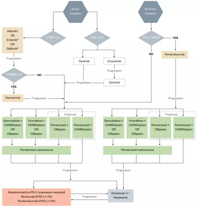
Figure 2: Advanced non-squamous cell NSCLC-SACT pathway (histology and molecular testing complete)
Treatment algorithm outlining lines of treatment available for adenocarcinoma cell non–small-cell lung cancer dependent on PD-L1 status and genetic mutations.Advanced non-squamous cell NSCLC-SACT pathway
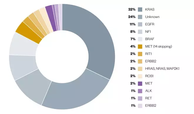
Figure 3: Genomic classification of adenocarcinoma subtypes
Pie chart illustrating percentages of varying genetic mutations in adenocarcinomas of the lung.
Immunotherapy
Using the immune system to treat cancer by inducing, enhancing or suppressing an immune response is not a novel concept. The first breakthrough in immunotherapy was not in the treatment of lung cancer, but in melanoma. From the identification of cytotoxic T-lymphocyte antigen 4 (CTLA-4)[38] to its function as an immune checkpoint inhibitor[39] and its blockade as a potential cancer treatment[40] , in 2011 the US Food and Drug Administration approved the monoclonal antibody ipilimumab for stage 4 melanoma. The monoclonal antibody PD-1 inhibitors were then identified with approval of pembrolizumab and nivolumab, and their application across multiple tumour sites, including renal and lung cancer.Checkpoint inhibitors
Immune checkpoints are biologically necessary to mediate immune response, thereby preventing detrimental inflammation and autoimmune diseases. Cancers by their very nature have disrupted cell synthesis pathways and, consequently, produce abnormal proteins that are immunogenic. Some cancers are able to ‘hijak’ immune checkpoints, leading to immune tolerance and further progression of the cancer. There are two main types of checkpoint inhibitors: those that effect PD-1 or PD-L1, and those effecting CTLA-4. Both operate at different stages of the immune response[41] . See this recent learning article for more information on immune checkpoint inhibitors in cancer: bit.ly/PJ-ICinhibitors .Mechanisms of action
CTLA-4 inhibitors
Cytotoxic T cells (CTLs) are activated by antigen presenting cells (via the major histocompatibility complex). For this activation to occur, there must be binding of the molecule CD28 on CTLs with a B7 molecule on the antigen presenting cell. When a CTL is activated, CTLA-4 (a further molecule on CTLs that has a higher binding affinity for B7) is able to out-compete CD28 for binding to B7. This inhibits further CTL activation and reduces the magnitude and duration of the CTL response. This has an important physiological function to reduce immune response once a pathogenic agent has been dealt with. This mechanism to reduce effectiveness of CTLs is utilised by cancer cells, which, by producing CTLA-4 on their cell surface, are able to block the co-activation of the CTLs. By inhibiting the CTLA-4 molecule on the CTL, this prevents the CTLs being ‘turned off’ and CTLs continue to destroy cancer cells (Figure 4)[42] . 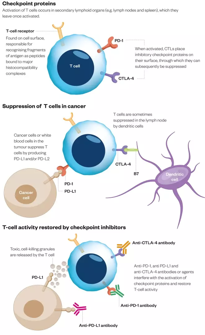
Figure 4: Checkpoint inhibitors mechanism of action
Diagram to show mechanism of action of PD-1/PD-L1 and CTLA-4 checkpoint inhibitors.
PD-1/PD-L1 inhibitors
PD-1 is found on tissue macrophages, while its ligands PD-L1 and PD-L2 are found on the surface of CTLs. The binding of PD-1 and PD-L1/2 is used biologically to suppress activity of CTLs once its role in fighting off infection is successful. The ligands PD-L1 and PD-L2 can be over-expressed by cancer cells and are a mechanism for cancers to evade the CTL anti-tumour response. PD-1 inhibitors nivolumab and pembrolizumab block the binding of PD-1 to both PD-L1 and PD-L2, while PD-L1 inhibitor atezolizumab blocks the binding of PD-1 to PD-L1 only (Figure 4). PD-1/PD-L1 inhibitors only reactivate CTLs that are already engaged in the anti-tumour response. This results in a more restricted T-cell reactivation with PD-1/PD-L1 blockade than with CTLA-4 blockade. This in part may explain the lower incidence of immune-related toxicities seen in PD-1/PD-L1 blockade compared with CTL1-4 blockade.Evidence in NSCLC for immunotherapy
Four landmark randomised phase III trials have demonstrated clinically significant benefit in advanced NSCLC patients treated with PD-1/PD-L1 inhibitors (nivolumab, pembrolizumab and atezolizumab) over docetaxel in the previously treated patient group[43],[44],[45],[46] (see Table 1).| Trial | Checkpoint inhibitor | Patient group | Endpoints |
|---|---|---|---|
| OS = overall survival; PD-L1 = programmed death receptor 1 ligand; PFS = progression-free survival | |||
| CheckMate-017 | Nivolumab | Squamous cell No PD-L1 expression necessary | OS 9.2 months vs. 6.0 months PFS 3.5 months vs. 2.8 months |
| CheckMate-057 | Nivolumab | Non-squamous cell No PD-L1 expression necessary | OS 12.2 months vs. 9.4 months PFS 2.3 months vs. 4.2 months |
| KeyNote 010 | Pembrolizumab | PD-L1-expressors only | OS 14·9 months vs. 8·2 months PFS 5·0 months vs. 4·1 months |
| OAK | Atezolizumab | Squamous and non-squamous cell No PD-L1 expression necessary | Dependent on histology and PD-L1 expression but overall survival improvement in all cohorts |
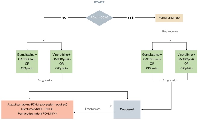
Figure 5: Advanced squamous cell NSCLC-SACT pathway (histology and molecular testing complete)
Treatment algorithm outlining lines of treatment available for squamous cell non–small-cell lung cancer dependent on PD-L1 status.
PD-L1 as a biomarker
There is debate as to whether PDL-1 is a useful tool for patient selection. Several trials show PFS and overall survival increase with higher expression of PD-L1 levels[43],[44],[45],[46] ; however, in the Checkmate-017 trial[43] it was found that in patients with squamous histology, PD-L1 was not predictive of response to nivolumab; there were patients who achieved durable benefit from PD-1/PD-L1 inhibitors despite having PD-L1-negative tumours. With regards to this uncertainty, NICE approved nivolumab in patients with squamous histology regardless of PD-L1 expression, but for non-squamous patients only those who have a PD-L1 score ≥1%. The results of the OAK trial with atezolizumab exacerbated this uncertainty, exhibiting responses in PD-L1 non-expressors both in the squamous and non-squamous group[46] . The reliability of the test for PD-L1 has also been drawn into question, as well as the cut-off point for PD-L1 expression. Different assays and cut-off points were applied to each drug during development and, as such, reproducibility of results is also questioned. The Blueprint PD-L1 IHC Assay Comparison Project investigated the analytical and clinical comparability of four PD-L1 IHC assays used in clinical trials. It was found that, despite three of the four assays tested being closely aligned, interchanging of assays and cut-offs could lead to misclassification of PD-L1 status in some patients[52] . Alternative biomarkers to PD-L1 expression are being investigated, such as total mutation burden, where higher levels of tumour mutation burden seem to correlate with an increased response to PD-1/PD-L1 inhibitors[53] .Side effects of PD-1/PD-L1 inhibitors
PD-1/PD-L1 inhibitors have clearly demonstrated a role for treating advanced NSCLC. The emerging challenge within the clinic is managing the distinctive spectrum of side effect profiles with these agents when compared with conventional chemotherapy. Oncologists and oncology pharmacists are well versed in managing chemotherapy-related adverse events (e.g. neutropenic sepsis); however, non-specialists must also be aware of the immune-related adverse events (irAEs) that develop from PD-1/PD-L1 inhibitor use. In general, irAEs occur quite early, mostly within weeks to three months after initiation of immune checkpoint blockers. However, the first onset of irAEs has been documented after up to two years of therapy and even up to a year after completion/cessation of therapy[43],[44],[45],[46] . The most commonly seen irAEs include dermatologic, gastrointestinal, hepatic and endocrine complications. IrAEs are believed to arise from general immunologic enhancement, and so temporary immunosuppression with corticosteroids, tumour necrosis factor-alpha antagonists, or other autoimmune inhibitory agents can be an effective treatment in most cases (see Treatment of irAEs). In general, irAEs appear to be less severe in PD-1/PDL-1 inhibitors than in CTLA-4 inhibitors. The most frequently reported adverse event across single drug studies is fatigue for 16–37% of patients on PD-1 inhibitors and 12–24% of patients on PD-L1 inhibitors[54] . Fatigue can be attributed to hypothyroidism in only a minority of patients (which is a separate common irAE associated with checkpoint inhibitors).Patient selection
Before initiation of PD-1/PD-L1 inhibitors, patients should be assessed for the likelihood of developing irAEs, including patient history, especially previous autoimmune disease and baseline laboratory tests. Patients who have a history of autoimmune disease (e.g. inflammatory bowel disease or rheumatoid arthritis) are at risk for worsening of their autoimmune disease while on checkpoint inhibitors. There are very little data to support treatment of this patient group, as the autoimmune population were excluded from the pivotal immunotherapy trials. Results from a retrospective review seem to show around a 30% chance of instigation of a symptom flare in pre-existing conditions and similar response rates in the treated cancer to the normal population[55] . These episodes appeared to respond to standard rescue therapy. However, this is an area that requires prospective research and any intent to treat at present should be undertaken with extreme caution and consideration of the potential risk versus benefit. Patients should be informed of the potential interactions regarding irAEs before treatment initiation. The appropriate specialist in the patient’s autoimmune disease should be contacted and involved prior to any therapy commencing. The patient should be aware who to contact at the earliest suspicion of irAEs and that this must be done as a matter of urgency.Interactions of immunotherapies
As monoclonal antibodies are not metabolised by cytochrome P450 enzymes or other drug-metabolising enzymes, inhibition or induction of these enzymes by co-administered medicines is not thought to affect the pharmacokinetics of the immunotherapies. The use of systemic corticosteroids or immuno-suppressants before starting immunotherapy should be avoided because of their potential interference with the pharmacodynamic activity and efficacy of immunotherapies. However, systemic corticosteroids or other immunosuppressants can be used after starting immunotherapies to treat irAEs[56],[57] .Treatment of irAEs
In general, treatment of moderate or severe irAEs requires interruption of the checkpoint inhibitor and the use of corticosteroid immunosuppression. Treatment is based upon the severity of the observed toxicity. For patients with grade 2 (moderate) irAEs, treatment with the checkpoint inhibitor should be withheld and should not be resumed until symptoms or toxicity is grade 1 or less. Corticosteroids, such as prednisolone 60mg once daily, should be started if symptoms do not resolve. For patients experiencing grade 3 or 4 (severe or life-threatening) irAEs, the checkpoint inhibitor should be permanently discontinued. High doses of corticosteroids such as prednisolone or methylprednisolone should be given (intravenous methylprednisolone 1–2mg/kg; prednisolone 1mg/kg max 60mg once daily). When symptoms subside to grade 1 or less, steroids can be gradually tapered; however, if reduced too quickly, then failure of recovery can commonly occur. Importantly, so far there is no evidence that the long-term clinical outcome of patients on PD-1/PD-L1 inhibitors are affected by the use of immunosuppressive agents for the management of irAEs[58] . However, the use of high-dose steroids for a prolonged period of time may be required and, as such, long-term side effects of steroid use may be expected in these patients.Pharmacist’s role
Pharmacists may be involved in patient care at a variety of stages throughout a patient’s care pathway. Smoking cessation advice provided by both primary and secondary care pharmacists plays a pivotal role in preventing lung cancer occurring in the first instance. Community pharmacists also have an important role to play in recognising red flags (see Box 1) and signposting for further investigation. Pharmacists involved in the care of respiratory patients, particularly patients with smoking-related respiratory disease, need to be observant of clinical changes in this high-risk patient group. In patients with a confirmed lung cancer diagnosis who are fit for surgery, pharmacists have a role in optimising pre- and post-surgical management. Where surgery or radiotherapy is not possible and patients require SACT, oncology pharmacists are well placed to explain the possible benefits of treatments to patients, balanced with the side effects of treatments and providing ongoing information on treatment. Pharmacists placed in A&Es and medical assessment units need to be knowledgeable of the differences between chemotherapy, targeted therapies and immunotherapies, and the management of toxicities of each modality. Furthermore, palliative care pharmacists substantially impact a person with lung cancer’s quality of life by providing supportive care in the last months to years of life. By working within the part of the multi-disciplinary team that surrounds the patient, pharmacists employed in various sectors can and do much to improve life for people living with lung cancer.- Having a cough for over three weeks (repeated requests for cough medicines of antibiotics);
- Being short of breath (increase in inhaler usage);
- Coughing up phlegm (sputum) with signs of blood in it;
- An ache or pain in the chest or shoulder (repeated requests for over-the-counter painkillers);
- Loss of appetite;
- Tiredness (fatigue);
- Losing weight;
- Hoarse voice;
- Difficulty swallowing;
- Pain and discomfort under the ribs, in the chest or shoulder;
- Finger clubbing;
- Facial or neck swelling.
Future challenges
The main advances in lung cancer in the past decade has been in the NSCLC advanced setting, with the curative options for NSCLC remaining largely unchanged (see Figure 6). However, the recent PFS benefit of durvalumab when compared with placebo as maintenance treatment after chemo-radiation for stage 3 disease is the first step into the adjuvant setting for PD-1/PD-L1 inhibitors[59] . Durvalumab is currently only available for use in the NHS via an Early Access to Medicines Scheme (EAMS). In SCLC, there is also an expanding portfolio of evidence of checkpoint inhibitors (STIMULI and CheckMate 451)[60],[61] in a group of patients where treatments and outcomes have varied little for decades. There is currently great interest in the interactions between the immune system, immunotherapy and radiation therapy, with questions raised around the potential immunogenic action of radiation therapy energising the immune system, leading to synergistic and abscopal effects. The success of targeted agents in small subgroups of patients highlights the need to test for molecular alterations and for investigation of further targets. The use of biomarkers in basket and umbrella trials should be encouraged to grant timely access to new active agents to patients, with the UK at the forefront with studies such as Matrix and TRACERx. PD-1/PD-L1 inhibitors can produce durable benefits with long-term survival rates and favourable safety profiles, meaning their use is now standard practice. As such, there is now a drive to use PD-1/PD-1s in combination with chemotherapy, targeted agents, radiation and with other immune oncology agents. However, careful consideration must be taken to manage substantial toxicities, as well the certain financial burden these strategies will bring. The future challenge will be introducing the new treatments into clinical practice and establishing real-world efficacy, managing side effects and monitoring for the development of as yet unseen long-term toxicity, along with diversifying into the rarer subtypes of thoracic cancer (e.g. SCLC and mesothelioma). 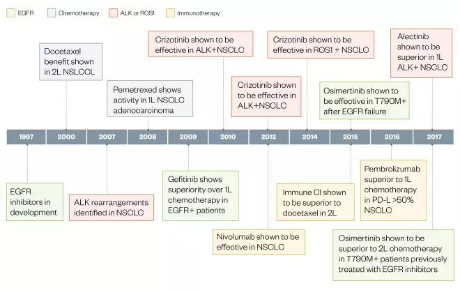
Figure 6: Developments in non-small-cell lung cancer (1997-2017)
Timeline illustration of developments in non–small cell lung cancer over the past decade.
1L: first line; 2L: second line; ALK: anaplastic lymphoma kinase; EGFR: epidermal growth factor receptor; NSCLC: non-small-cell lung cancer; TKI: tyrosine kinase inhibitor.
References
[1] Cancer Research UK. Lung cancer statistics. Available at: http://www.cancerresearchuk.org/health-professional/cancer-statistics/statistics-by-cancer-type/lung-cancer? (accessed June 2018)
[2] National Institute for Health and Care Excellence. Lung cancer overview. Available at: https://pathways.nice.org.uk/pathways/lung-cancer (accessed June 2018)
[3] Kaseda S, Aoki T, Hangai N & Shimizu K. Better pulmonary function and prognosis with video-assisted thoracic surgery than with thoracotomy. Ann Thorac Surg 2000;70(5):1644–1646. doi: 10.1016/S0003-4975(00)01909-3
[4] Yim AP. VATS major pulmonary resection revisited–controversies, techniques, and results. Ann Thorac Surg 2002;74(2):615–623. doi: 10.1016/S0003-4975(02)03579-8
[5] Berry MF. Pulmonary artery bleeding during video-assisted thoracoscopic surgery: intraoperative bleeding and control. Thorac Surg Clin 2015;25(3):239–247. doi: 10.1016/j.thorsurg.2015.04.007
[6] Abreu CECV, Ferreira PPR, de Moraes FY et al. Stereotactic body radiotherapy in lung cancer: an update. J Brasileiro de Pneumologia 2015;41(4):376–387. doi: 10.1590/S1806-37132015000000034
[7] Curran WJ, Jr, Paulus R, Langer CJ et al. Sequential vs. concurrent chemoradiation for stage III non-small-cell lung cancer: randomized phase III trial RTOG 9410. J Natl Cancer Inst 2011;103:1452–1460. doi: 10.1093/jnci/djr325
[8] Saunders M, Dische S, Barrett A et al. Continuous hyperfractionated accelerated radiotherapy (CHART) versus conventional radiotherapy in non-small-cell lung cancer: a randomised multicentre trial. CHART Steering Committee. Lancet 1997;350:161–165. doi: 10.1016/S0140-6736(97)06305-8
[9] Bradley DJ, Paulus R, Komaki R et al. A randomized phase III comparison of standard-dose (60 Gy) versus high-dose (74 Gy) conformal chemoradiotherapy with or without cetuximab for stage III non-small cell lung cancer. Results on radiation dose in RTOG 0617. J Clin Oncol 2013;31: abstr 7501. doi: 10.1200/jco.2013.31.15_suppl.7501
[10] National Comprehensive Cancer Network. Non-small-cell lung cancer NCCN guidelines. 2018. Available at: https://www.nccn.org/patients/guidelines/lung-nsclc/ (accessed June 2018)
[11] Royal College of Physicians. National Lung Cancer Audit 2017 (for the audit period 2016). Available at: www.rcplondon.ac.uk/nlca (accessed June 2018)
[12] Schiller JH, Harrington D, Belani CP et al. Comparison of four chemotherapy regimens for advanced non–small-cell lung cancer. N Engl J Med 2002;346:92–98. doi: 10.1056/NEJMoa011954
[13] Scagliotti GV, Parikh P, von Pawel J et al. Phase III study comparing cisplatin plus gemcitabine with cisplatin plus pemetrexed in chemotherapy-naive patients with advanced-stage non–small-cell lung cancer. J Clin Oncol 2008;26:3543–3551. doi: 10.1200/JCO.2007.15.0375
[14] Lynch TJ, Bell DW, Sordella R et al. Activating mutations in the epidermal growth factor receptor underlying responsiveness of non–small-cell lung cancer to gefitinib. N Engl J Med 2004;350:2129–2139. doi: 10.1056/NEJMoa040938
[15] Mitsudomi T, Morita S, Yatabe Y et al. Gefitinib versus cisplatin plus docetaxel in patients with non-small-cell lung cancer harbouring mutations of the epidermal growth factor receptor (WJTOG3405): an open label, randomised phase III trial. Lancet Oncol 2010;11:121–128. doi: 10.1016/S1470-2045(09)70364-X
[16] Rosell R, Carcereny E, Gervais R et al. Erlotinib versus standard chemotherapy as first-line treatment for European patients with advanced EGFR mutation-positive non-small-cell lung cancer (EURTAC): a multicentre, open-label, randomised phase III trial. Lancet Oncol 2012;13:239–246. doi: 10.1016/S1470-2045(11)70393-X
[17] Wu Y-L, Zhou C, Hu C-P et al. Afatinib versus cisplatin plus gemcitabine for first-line treatment of asian patients with advanced non-small-cell lung cancer harbouring EGFR mutations (LUX-Lung 6): an open-label, randomised phase 3 trial. Lancet Oncol 2014;15:213–222. doi: 10.1016/S1470-2045(13)70604-1
[18] National Institute for Health and Care Excellence. Gefitinib the first-line treatment of locally advanced or metastatic non-small-cell lung cancer (TA192). 2010. Available at: https://www.nice.org.uk/guidance/ta192 (accessed June 2018)
[19] National Institute for Health and Care Excellence. Erlotinib for the first-line treatment of locally advanced or metastatic EGFR-TK mutation-positive non-small-cell lung cancer (TA258). 2012. Available at: https://www.nice.org.uk/guidance/ta258 (accessed June 2018)
[20] National Institute for Health and Care Excellence. Afatinib for treating epidermal growth factor receptor mutation-positive locally advanced or metastatic non-small-cell lung cancer (TA310). 2014. Available at: https://www.nice.org.uk/guidance/ta310 (accessed June 2018)
[21] Kohno T, Nakaoku T, Tsuta K et al. Beyond ALK-RET, ROS1 and other oncogene fusions in lung cancer. Transl Lung Cancer Res 2015;4:156–164. doi: 10.3978/j.issn.2218-6751.2014.11.11
[22] Yu HA, Arcila ME, Rekhtman N et al. Analysis of tumor specimens at the time of acquired resistance to EGFR-TKI therapy in 155 patients with EGFR-mutant lung cancers. Clin Cancer Res 2013;19:2240–2247. doi: 10.1158/1078-0432.CCR-12-2246
[23] Mok TS, Y-L W, Ahn M-J, Garassino MC et al. Osimertinib or platinum-pemetrexed in EGFR T790M-positive lung cancer. N Engl J Med 2016;376:629–640. doi: 10.1056/NEJMoa1612674
[24] National Institute for Health and Care Excellence. Osimertinib for treating locally advanced or metastatic EGFR T790M mutation-positive non-small-cell lung cancer (TA416). 2016. Available at: https://www.nice.org.uk/guidance/ta416 (accessed June 2018)
[25] Reckamp KL, Melnikova VO, Karlovich C et al. A highly sensitive and quantitative test platform for detection of NSCLC EGFR mutations in urine and plasma. J Thorac Oncol 2016;11:1690–1700. doi: 10.1016/j.jtho.2016.05.035
[26] Ramalingam SS. Osimertinib vs standard of care (SoC) EGFR-TKI as first-line therapy in patients (pts) with EGFRm advanced NSCLC: FLAURA. Ann Oncol 2017;28(5):v605–v649. doi: 10.1093/annonc/mdx440.050
[27] National Institute for Health and Care Excellence. Crizotinib for untreated anaplastic lymphoma kinase-positive advanced non-small-cell lung cancer (TA406). 2016. Available at: https://www.nice.org.uk/guidance/ta406 (accessed June 2018)
[28] National Institute for Health and Care Excellence. Ceritinib for untreated ALK-positive non-small-cell lung cancer (TA500). 2018. Available at: https://www.nice.org.uk/guidance/ta500 (accessed June 2018)
[29] National Institute for Health and Care Excellence. Alectinib for untreated anaplastic lymphoma kinase-positive advanced non-small-cell lung cancer (in development [GID-TA10206]). Available at: https://www.nice.org.uk/guidance/indevelopment/gid-ta10206 (accessed June 2018)
[30] Shaw AT, Kim D-W, Nakagawa K et al. Crizotinib versus chemotherapy in advanced ALK–positive lung cancer. N Engl J Med 2013;368:2385–2394. doi: 10.1056/NEJMoa1214886
[31] Solomon BJ, Mok T, Kim D-W et al. First-line crizotinib versus chemotherapy in ALK–positive lung cancer. N Engl J Med 2014;371:2167–2177. doi: 10.1056/NEJMoa1408440
[32] Crinò L, Ahn M-J, De Marinis F et al. Multicenter phase II study of whole-body and intracranial activity with ceritinib in patients with ALK–rearranged non–small-cell lung cancer previously treated with chemotherapy and crizotinib: results from ASCEND-2. J Clin Oncol 2016;34:2866–2873. doi: 10.1200/JCO.2015.65.5936
[33] Soria J-C, Tan DSW, Chiari R et al. First-line ceritinib versus platinum-based chemotherapy in advanced ALK-rearranged non-small-cell lung cancer (ASCEND-4): a randomised, open-label, phase 3 study. Lancet 2017;389:917–929. doi: 10.1016/S0140-6736(17)30123-X
[34] Peters S, Camidge DR, Shaw AT et al. Alectinib versus crizotinib in untreated ALK–positive non–small-cell lung cancer. N Engl J Med 2017;377:829–838. doi: 10.1056/NEJMoa1704795
[35] Shaw AT, Ou S-HI, Bang Y-J et al. Crizotinib in ROS1–rearranged non-small-cell lung cancer. N Engl J Med 2014;371:1963–1971. doi: 10.1056/NEJMoa1406766
[36] Toschi L, Rossi S, Finocchiaro G et al. Non-small cell lung cancer treatment (r)evolution: ten years of advances and more to come. Ecancermedicalscience 2017;11:787. doi: 10.3332/ecancer.2017.787
[37] O’Neill A, Jagannathan JP & Ramaiva NH. Evolving cancer classification in the era of personalized medicine: a primer for radiologists. Korean J Radiol 2017;18(1):6–17. doi: 10.3348/kjr.2017.18.1.6
[38] Brunet JF, Denizot F, Luciani MF et al. A new member of the immunoglobulin superfamily — CTLA-4. Nature 1987;328:267–270. doi: 10.1038/328267a0
[39] Krummel MF & Allison JP. CD28 and CTLA-4 have opposing effects on the response of T cells to stimulation. J Exp Med 1995;182:459–465. Available at: http://jem.rupress.org/content/182/2/459.long (accessed June 2018)
[40] Leach DR, Krummel MF & Allison JP. Enhancement of anti-tumor immunity by CTLA-4 blockade. Science 1996;271:1734–1736. doi: 10.1126/science.271.5256.1734
[41] Fife BT & Bluestone JA. Control of peripheral T-cell tolerance and autoimmunity via the CTLA-4 and PD-1 pathways. Immunol Rev 2008;224:166–182. doi: 10.1111/j.1600-065X.2008.00662.x
[42] Luke JJ & Ott PA. PD-1 pathway inhibitors: the next generation of immunotherapy for advanced melanoma. Oncotarget 2015;6(6):3479–3492. doi: 10.18632/oncotarget.2980
[43] Brahmer J, Reckamp KL, Baas P et al. Nivolumab versus docetaxel in advanced squamous-cell non-small-cell lung cancer. N Engl J Med 2015;373:123–135. doi: 10.1056/NEJMoa1504627
[44] Borghaei H, Paz-Ares L, Horn L et al. Nivolumab versus docetaxel in advanced nonsquamous non-small-cell lung cancer. N Engl J Med 2015;373:1627–1639. doi: 10.1056/NEJMoa1507643
[45] Herbst RS, Baas P, Kim D-W et al. Pembrolizumab versus docetaxel for previously treated, PD-L1-positive, advanced non-small-cell lung cancer (KEYNOTE-010): a randomised controlled trial. Lancet 2016;387:1540–1550. doi: 10.1016/S0140-6736(15)01281-7
[46] Rittmeyer A, Barlesi F, Waterkamp D et al. Atezolizumab versus docetaxel in patients with previously treated non-small-cell lung cancer (OAK): a phase 3, open-label, multicentre randomised controlled trial. Lancet 2017;389:255–265. doi: 10.1016/S0140-6736(16)32517-X
[47] National Institute for Health and Care Excellence. Nivolumab for previously treated squamous non-small-cell lung cancer (TA483). 2017. Available at: https://www.nice.org.uk/guidance/ta483 (accessed June 2018)
[48] National Institute for Health and Care Excellence. Nivolumab for previously treated non-squamous non-small-cell lung cancer (TA484). 2017. Available at: https://www.nice.org.uk/guidance/ta484 (accessed June 2018)
[49] National Institute for Health and Care Excellence. Pembrolizumab for treating PD-L1-positive non-small-cell lung cancer after chemotherapy (TA428). 2017. Available at: https://www.nice.org.uk/guidance/ta428 (accessed June 2018)
[50] National Institute for Health and Care Excellence. Atezolizumab for treating non-small-cell lung cancer after platinum-based chemotherapy [ID970]. 2018. Available at: https://www.nice.org.uk/guidance/indevelopment/gid-ta10108 (accessed June 2018)
[51] Reck M, D Rodriguez-Abreu, Robinson AG et al. Pembrolizumab versus chemotherapy for PD-L1 positive non-small-cell lung cancer (KEYNOTE-024). N Engl J Med 2016;375:1823–1833. doi: 10.1056/NEJMoa1606774
[52] Hirsch FR, McElhinny A, Stanforth D et al. PD-L1 immunohistochemistry assays for lung cancer: results from phase 1 of the Blueprint PD-L1 IHC Assay Comparison Project. J Thorac Oncol 2017;12(2):208–222. doi: 10.1016/j.jtho.2016.11.2228
[53] Spigel DR, Schrock AB, Fabrizio D et al. Total mutation burden (TMB) in lung cancer (LC) and relationship with response to PD-1/PD-L1 targeted therapies. J Clin Oncol 2016;34:9017. doi: 10.1200/JCO.2016.34.15_suppl.9017
[54] Naidoo J, Page DB, Li BT et al. Toxicities of the anti-PD-1 and anti-PD-L1 immune checkpoint antibodies. Ann Oncol 2015;26:2375–2391. doi: 10.1093/annonc/mdv383
[55] Spencer KR & Mehnert JM. Immunotherapy in patients with active autoimmune disease. Available at: https://immunosym.org/daily-news/immunotherapy-patients-active-autoimmune-disease (accessed June 2018)
[56] Electronic Medicines Compendium. OPDIVO 10 mg/mL concentrate for solution for infusion. 2018. Available at: https://www.medicines.org.uk/emc/product/6888/smpc (accessed June 2018)
[57] Electronic Medicines Compendium. KEYTRUDA 25 mg/mL concentrate for solution for infusion. 2018. Available at: https://www.medicines.org.uk/emc/product/2498/smpc (accessed June 2018)
[58] Horvat TZ AdelNG, DangTO et al. Immune-related adverse events, need for systemic immunosuppression, and effects on survival and time to treatment failure in patients with melanoma treated with ipilimumab at Memorial Sloan Kettering Cancer Center. J Clin Oncol 2015;33:3193–3198. doi: 10.1200/JCO.2015.60.8448
[59] Antonia SJ, Villegas A, Daniel D et al. Durvalumab after chemoradiotherapy in stage III non-small-cell lung cancer. N Engl J Med. doi: 10.1056/NEJMoa1709937
[60] De Ruysscher D, Pujol J-L, Popat S et al. STIMULI: a randomised open-label phase II trial of consolidation with nivolumab and ipilimumab in limited-stage SCLC after standard of care chemo-radiotherapy conducted by ETOP and IFCT. Ann Oncol 2016;27(Suppl 6):1430TiP. doi: 10.1093/annonc/mdw389.08
[61] Ready N, Owonikoko TK, Postmus PE et al. CheckMate 451: A randomized, double-blind, phase III trial of nivolumab (nivo), nivo plus ipilimumab (ipi), or placebo as maintenance therapy in patients (pts) with extensive-stage disease small cell lung cancer (ED-SCLC) after first-line platinum-based doublet chemotherapy (PT-DC). J Clin Oncol 2016;34(15 Suppl):7000. doi: 10.1200/JCO.2016.34.15_suppl.TPS8579
