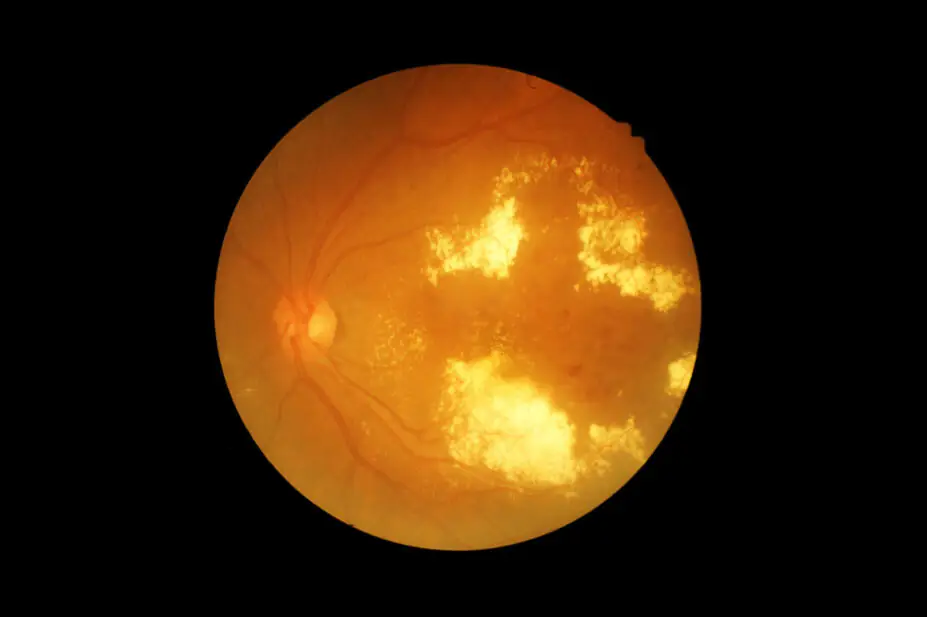
memorisz / Shutterstock.com
After reading this article, you should be able to:
- Describe the pathophysiology, risk factors, signs and symptoms of diabetic retinopathy;
- Identify how diabetic retinopathy is diagnosed through national screening and when referral to specialist services is required;
- Understand evidence-based pharmacological and non-pharmacological management used to manage diabetic retinopathy and its complications;
- Understand how treatment choices can affect patient burden and capacity in services;
- Demonstrate ways pharmacists can work with patients living with diabetic retinopathy to optimise outcomes.
Introduction
Diabetes affects around 4.5 million adults in the UK1. Diabetic retinopathy — a type of diabetic eye disease — is a microvascular complication of diabetes affecting retinal blood vessels. It’s a leading cause of visual impairment and blindness, particularly in working-age adults (i.e. those aged 20-65 years) with the condition, who have a 20-fold risk increase of blindness compared with individuals without diabetes2.
The UK prevalence of retinopathy in individuals with diabetes is estimated to be around 34%, with 12% classified as sight-threatening3. Higher rates of diabetic retinopathy are observed in men, people of South Asian and Afro-Caribbean ethnicity, as well as those from socioeconomically deprived backgrounds4.
Chronic hyperglycaemia is a key driver of retinopathy, while most patients with diabetes will develop retinopathy to some extent within 20 years of diagnosis5. People with both type 1 diabetes mellitus (T1DM) and type 2 diabetes mellitus (T2DM) are at risk.
Owing to later diagnosis, retinopathy is more common at the initial presentation of T2DM. However, the cumulative long-term risk is higher in people with T1DM (95% versus 60% over 15 years)2. Additional risk factors include a longer duration of disease, poor glycaemic control and diabetes-related comorbidities2. Other ocular conditions — including cataracts and glaucoma — are also more prevalent in people with diabetes2.
People with, or at risk of, diabetic retinopathy often experience substantial healthcare burden, which has a significant impact on those in work or education. This is because they are required to attend regular appointments across multiple health services and need lifelong management of their condition6.
Visual impairment resulting from diabetic retinopathy also impacts psychological wellbeing and the ability to complete everyday tasks such as driving and reading7. A national screening programme was established in 2003 to enable early detection and timely referral for treatment8, while National Institute for Health and Care Excellence (NICE) guidelines have been published to optimise management5.
Pharmacists play an important part in optimising patient outcomes. Contributions may include supporting lifestyle changes, improving glycaemic control (i.e. with and without medication), promoting attendance to regular eye screening appointments, assessing adherence to treatment and identifying ‘red flags’ that require urgent referral.
Risk factors
Anyone with T1DM or T2DM is at risk of developing diabetic retinopathy, with additional risk increased by the following:
- Poor glycaemic control (i.e. raised HbA1c);
- Duration of diabetes or late diagnosis;
- Hypertension;
- Dyslipidaemia;
- Smoking;
- Obesity;
- Diabetes-related comorbidities (e.g. nephropathy);
- Pregnancy with pre-existing diabetes;
- Ethnicity, as there is a higher risk in South Asian and Afro-Caribbean populations;
- Male gender;
- Family history2,9.
Pathophysiology
The retina has a high metabolic demand, with oxygen and nutrients supplied through the central retinal artery and its capillaries10.
Persistently raised blood glucose levels can lead to biochemical and structural changes to the retinal vasculature such as oxidative stress, inflammation and the accumulation of advanced glycation end-products formed through the non-enzymatic glycation of proteins and lipids10–12.
Capillaries become more fragile, microaneurysms form and vascular permeability increases12. This results in the leakage of fluid, lipids and blood into retinal tissue and can cause diabetic macular oedema (DMO), with the macula being the part of the retina responsible for central vision. This leakage distorts the layers of the retina, which subsequently affects vision11,12.
Additionally, in response to vascular damage, angiogenic factors, such as vascular endothelial growth factor (VEGF), are released, which stimulates the formation of new, fragile and highly permeable blood vessels prone to haemorrhage12.
This is known as proliferative diabetic retinopathy (PDR), which can lead to further sight-threatening complications, including vitreous haemorrhage, fibrosis (i.e. scar formation), retinal detachment and neovascularisation of the iris, which may cause glaucoma (see Figure 110–12).
Figure 1: Diabetic retinopathy compared with healthy retina
Screening considerations
Early diabetic retinopathy is frequently asymptomatic and usually detected through routine screening2,11. Patients with more advanced disease may present with reduced, blurred, fluctuating vision or floaters2.
Early detection of diabetic eye disease is vital. The more prompt the treatment, the more outcomes are improved for patients. The NHS Diabetic Eye Screening Programme (DES) has been established by the UK National Screening Committee (UK NSC) to provide screening in anyone with T1DM and T2DM aged 12 years and over, which is identified through GP records8.
Each nation within the UK has its own screening policy based on these recommendations — although, there may be slight differences between them. For example, Scotland offers six-monthly screening in some high-risk cases13.
Invitations for screening are sent to eligible patients annually; however, if no signs of diabetic retinopathy are seen on two consecutive visits, this will reduce to once every two years as the risk of developing sight-threatening disease is lower14,15. Attendance is not mandatory but should be encouraged8. Those not wishing to attend regularly may request that invitations be deferred for up to three years16.
Appointments and diagnosis
The diabetic eye screening appointment involves the following:
- Measurement of patient’s visual acuity in each eye against a Snellen chart or equivalent;
- Instillation of dilating eye drops such as phenylephrine or tropicamide;
- Digital imaging taken of each retina using specialist equipment16.
Patients should be advised not to drive themselves to appointments as their vision may be blurry for up to four hours afterwards, while contact details should be provided for reporting unexpected adverse events17.
Patients who must avoid dilating eye drops — for example, if patients have acute angle-closure glaucoma — may have images taken without dilation. However, this should be noted as the quality of imaging is likely to be poorer17. If images cannot be taken successfully, referral for further assessment should be made18.
The administration of dilating eye drops by both registered and non-registered healthcare professionals during the screening process falls within the Medicines Act 1968 and may be permitted without the need for a prescription or patient group direction (PGD) — provided the eye drops are obtained within legislation17.
Images are graded against national standards to classify the extent of retinopathy and maculopathy (i.e. the presence of macular oedema) into ‘referable’ and ‘non-referable’ disease (see Table 12). Images of the retina are reviewed for presence of microaneurysms, haemorrhages or other exudates from leaking vessels, increased tortuosity of the retinal vasculature and the presence of neovascularisation.
Table 1: Grading of diabetic retinopathy
If referral to secondary care is made, further assessment and imaging may be undertaken, including:
- Slit-lamp and fundus examination of the eyes;
- Fundus fluorescein angiography, where fluorescein dye is injected intravenously and images taken of the retinal vasculature to identify venous haemorrhages, occlusions or neovascularisation;
- OCT, which shows a cross-section image of the retinal layers and is used to measure the thickness of the macula and monitor presence of oedema or structural changes2.
Pharmacists may encourage patients to attend regular eye screening and hospital appointments during structured medicine reviews or through health promotion campaigns involving diabetes care.
For example, ask: “Have you attended your diabetic eye screening appointment recently?” If not, discuss that retinopathy may develop or worsen without symptoms, as well as that early detection and treatment reduces the long-term risk of visual impairment. Pharmacists may also signpost patients to seek urgent advice if ‘red flag’ ocular symptoms are present. Local optometrists may offer urgent assessment under the Minor Eye Conditions Service (MECS). Otherwise, referral to the local A&E or eye casualty unit, if available, should be made (see Box)19.
Box: Ocular ‘red flags’ requiring urgent assessment
- Sudden loss of vision or ‘floaters’ obscuring vision;
- Eye pain, especially with headache and nausea/vomiting;
- Sensitivity to light;
- Mismatching pupil sizes;
- Blood or pus seen in the eye;
- Trauma to, or foreign object in, the eye19,20.
Diabetic retinopathy management
In 2024, NICE published guidelines on the management and monitoring of patients with diabetic retinopathy5. These guidelines recommend that all clinicians involved in patients’ diabetes care support them to optimise long-term control of their condition to reduce the impact of diabetic retinopathy5.
Non-pharmaceutical management in primary care
Lifestyle changes related to smoking, weight and exercise should be recommended2. Tight control of both HbA1c and blood pressure has been shown to reduce the incidence of microvascular disease, including retinopathy. A reduction in HbA1c from 9% to 7.2% in people with T1DM was shown to reduce the incidence of retinopathy by 76%2,21.
The results of a Cochrane review, published in 2023, demonstrated that interventions to control blood pressure reduced the five-year incidence of retinopathy; although, they did not significantly slow the progression of disease unless patients were hypertensive at presentation22. Off-label use of fenofibrate may reverse diabetes-induced retinal effects and may delay the progression of retinopathy in patients with T2DM. However, no evidence currently exists in T1DM23.
It should also be noted that there are concerns that a rapid reduction in HbA1c (e.g. with medication) can lead to the worsening of diabetic retinopathy and vision loss in the short term2,21. However, evidence reviewed by NICE did not demonstrate this — although it was of poor quality. It is therefore recommended that the patient’s ophthalmologist is made aware of any change to treatment that may lead to a rapid change in HbA1c, to allow appropriate assessment and monitoring5. Recent treatment developments, including GLP-1 inhibitors, require ongoing surveillance owing to evidence suggesting they may lead to early worsening of retinopathy24.
Pharmacists can educate people with diabetes on the risk of retinopathy and provide advice on optimising glycaemic control with or without medication. This may include support with quitting smoking, monitoring weight, blood pressure and blood glucose, as well as tailoring advice accordingly.
Management in secondary care
Monitoring and treatment of diabetic retinopathy is managed in secondary care by medical retina specialists. In patients with bilateral disease, eyes should be monitored and treated separately as they may show different stages of retinopathy5.
Even if one eye is affected more severely, treating both is important to help reduce progression of disease in either eye, which could have significant consequences5. Non-proliferative diabetic retinopathy (NPDR) should be monitored every 6–12 months if moderate, or every 3 months if severe, to detect progression to PDR or DMO early5. Evidence for active treatment of NPDR is currently lacking5.
PDR treatment
Treatment options for PDR include laser treatment (i.e. pan-retinal photocoagulation [PRP]) or injection of biologic agents targeting VEGF into the vitreous cavity of the eye (i.e. intravitreal route).
PRP involves using laser to burn areas of neovascularisation and haemorrhage, reducing fluid leakage into the retina. It does not improve vision by itself but can delay progression11. Anti-VEGF treatments are usually offered if PDR remains despite a complete course of PRP, which work by inhibiting VEGF and stopping the growth of new vessels5.
Only one anti-VEGF agent — ranibizumab — is currently licensed for patients with diabetic retinopathy without macular oedema; although, other medications may be used off-label depending on local commissioning agreements5.
If significant vitreous haemorrhage (i.e. not clearing within three months) or retinal detachment is present, removal of the vitreous gel and temporary replacement with gas or silicone (i.e. vitrectomy) may help. Vitreous gel will naturally turn over and replace itself over time5.
DMO treatment
Treatment options for DMO include intravitreal injection of anti-VEGF agents or steroid implants, or laser treatment to the macula5.
A macular laser aimed at areas of oedema rather than new vessels is recommended where oedema is not affecting central vision or where the patient’s visual is still good5.
Anti-VEGF agents
Where central vision is threatened and central retinal thickness (CRT) is larger than 400 microns — measured by OCT — anti-VEGF agents are recommended25. However, health inequalities may be avoided by offering treatment earlier to patient populations with naturally thinner retinas who may take longer to reach the CRT threshold. For example, those of South Asian and Afro-Caribbean descent, especially women5.
Anti-VEGF agents require initial loading — monthly injections for three to five months depending on the drug — followed by regimes tailored to the initial response to treatment25. This may be at fixed intervals or following a ‘treat and extend’ protocol where dosing intervals are increased between each injection until no further response is seen25.
Treatment should stop if there is significant worsening of oedema despite treatment or if irreversible damage occurs25. Newer agents, including high-dose aflibercept (8 mg) or faricimab, may offer longer dosing intervals of up to 16 weeks, which may help to reduce capacity issues in busy injection clinics and treatment burden for patients25.
Lower-cost, therapeutically equivalent biosimilar agents are available for ranibizumab and aflibercept (2 mg), but these may not achieve the same treatment intervals as newer agents25.
Overall, a balance between lower-cost and longer-acting agents needs to be found to optimise cost-effectiveness without overburdening services. NHS England has produced guidance to support both commissioners and ophthalmology services25.
Potential risks of intravitreal injections include endophthalmitis (i.e. an infection inside the eye), intraocular inflammation, retinal detachment and cataract. Injections should be administered only by trained healthcare professionals26.
Anti-VEGF agents carry a theoretical risk of systemic effects, including non-ocular bleeding and arterial thromboembolic events. Treatment should be withheld in patients with a recent history of stroke, transient ischaemic attack or myocardial infarction26. There is limited evidence for their use during pregnancy26.
Intravitreal corticosteroids
Intravitreal steroid implants offer an alternative where anti-VEGF treatment is contraindicated. This can include during pregnancy or where less frequent administration may be beneficial, if an individual has dementia or learning difficulties, for example. These implants release steroids slowly over time.
Dexamethasone is licensed for administration every six months, although in practice four-monthly administration is used owing to early loss of effect25. Fluocinolone implants release the drug over three years, although rescue treatment with anti-VEGF or dexamethasone is often required, which reduces cost-effectiveness25,27.
Additional risks for steroids can include cataract formation and raised intraocular pressure, which requires close monitoring27.
A summary of the available treatments for PDR and DMO can be seen in Table 24.
Table 2: Treatments recommended for diabetic retinopathy, as per NICE guidance
The pharmacist’s role
Pharmacists in secondary care settings may provide advice to patients regarding risks and benefits of intravitreal injections, as patients often report high anxiety levels and lack of understanding around intravitreal injections, especially before their first dose28.
They can also support services to switch treatments to biosimilar agents, where appropriate, to improve cost-effectiveness.
Best practice for pharmacists
Primary care
- Familiarise yourself with the eye services that are available locally. These may include eye casualty units, NHS Diabetic Eye Screening Programme services and optometrists that offer the minor eye conditions service. Building professional links with a local optometrist will likely benefit both services;
- Identify where diabetes is poorly controlled or risk factors could be modified and provide support to patients to optimise control. Signpost to support groups or health apps to help patients manage their condition;
- Ask patients how they manage their condition and if they attend their screening or hospital appointments. Identify barriers to attendance or adherence to treatment and discuss these with patients.
Secondary care
- Monitor the use of high-cost anti-vascular endothelial growth factor and steroid injections in your service against NHS England guidance to maximise cost-effectiveness. Identify barriers to switching to biosimilar agents and provide education to clinicians and patients to help address these;
- Support patients to better understand their injection treatment.
AI statement
The authors used Microsoft Co-Pilot for proofreading purposes prior to submission.
Disclosures
- Alimera — consultancy work;
- Roche — consultancy work and attendance at conferences.
- 1.IDF Diabetes Atlas 2025. International Diabetes Federation. https://diabetesatlas.org/resources/idf-diabetes-atlas-2025/
- 2.Denniston AKO, Murray PI, eds. Medical retina. Oxford Handbook of Ophthalmology. Published online March 2018:568-661. doi:10.1093/med/9780198804550.003.0013
- 3.Haider S, Thayakaran R, Subramanian A, et al. Disease burden of diabetes, diabetic retinopathy and their future projections in the UK: cross-sectional analyses of a primary care database. BMJ Open. 2021;11(7):e050058. doi:10.1136/bmjopen-2021-050058
- 4.Diabetic eye disease: A UK Incidence and Prevalence Study. The Royal National Institute of Blind People. 2017. https://media.rnib.org.uk/documents/Diabetic_eye_disease._A_UK_Incidence_and_Prevalence_Study_-_Full_report.pdf
- 5.Diabetic retinopathy: Management and Monitoring. The National Institute for Health and Care Excellence . 2024. https://www.nice.org.uk/guidance/NG242
- 6.Sivaprasad S, Oyetunde S. Impact of injection therapy on retinal patients with diabetic macular edema or retinal vein occlusion. OPTH. Published online May 2016:939. doi:10.2147/opth.s100168
- 7.Cooper OAE, Taylor DJ, Crabb DP, Sim DA, McBain H. Psychological, social and everyday visual impact of diabetic macular oedema and diabetic retinopathy: a systematic review. Diabet Med. 2019;37(6):924-933. doi:10.1111/dme.14125
- 8.Diabetic eye screening: programme overview. Public Health England . 2014. https://www.gov.uk/guidance/diabetic-eye-screening-programme-overview
- 9.Perais J, Agarwal R, Evans JR, et al. Prognostic factors for the development and progression of proliferative diabetic retinopathy in people with diabetic retinopathy. Cochrane Database of Systematic Reviews. 2023;2023(2). doi:10.1002/14651858.cd013775.pub2
- 10.Diabetic retinopathy symptoms and treatment. NHS Inform . https://www.nhsinform.scot/illnesses-and-conditions/diabetes/diabetic-retinopathy
- 11.Diabetic retinopathy — diabetes and eye problems. Diabetes UK. 2017. https://www.diabetes.org.uk/about-diabetes/looking-after-diabetes/complications/retinopathy
- 12.Gomułka K, Ruta M. The Role of Inflammation and Therapeutic Concepts in Diabetic Retinopathy—A Short Review. IJMS. 2023;24(2):1024. doi:10.3390/ijms24021024
- 13.Screening programmes across the UK. UK National Screening Committee. 2021. https://www.gov.uk/guidance/screening-programmes-across-the-uk
- 14.Leese GP, Stratton IM, Land M, et al. Progression of Diabetes Retinal Status Within Community Screening Programs and Potential Implications for Screening Intervals. Diabetes Care. 2014;38(3):488-494. doi:10.2337/dc14-1778
- 15.Diabetic Retinopathy . UK National Screening Committee . https://view-health-screening-recommendations.service.gov.uk/diabetic-retinopathy/
- 16.Diabetic eye screening. NHS. 2017. https://www.nhs.uk/tests-and-treatments/diabetic-eye-screening/
- 17.Use of mydriatic eye drops in the NHS diabetic eye screening programme. UK government . 2025. https://www.gov.uk/government/publications/nhs-diabetic-eye-screening-use-of-eye-drops/use-of-mydriatic-eye-drops-in-the-nhs-diabetic-eye-screening-programme
- 18.Diabetic eye screening: guidance when adequate images cannot be taken. UK government . 2026. https://www.gov.uk/government/publications/diabetic-eye-screening-pathway-for-images-and-where-images-cannot-be-taken/diabetic-eye-screening-guidance-when-adequate-images-cannot-be-taken
- 19.Minor Eye Conditions Service (MECS) covers minor eye problems. NHS Primary Eyecare. https://primaryeyecare.co.uk/services/minor-eye-conditions-service/
- 20.Clinical Knowledge Summary: Red eye. The National Institute for Health and Care Excellence . 2026. https://cks.nice.org.uk/topics/red-eye/
- 21.The Effect of Intensive Treatment of Diabetes on the Development and Progression of Long-Term Complications in Insulin-Dependent Diabetes Mellitus. N Engl J Med. 1993;329(14):977-986. doi:10.1056/nejm199309303291401
- 22.Do DV, Han G, Abariga SA, Sleilati G, Vedula SS, Hawkins BS. Blood pressure control for diabetic retinopathy. Cochrane Database of Systematic Reviews. 2023;2023(3). doi:10.1002/14651858.cd006127.pub3
- 23.Kataoka SY, Lois N, Kawano S, Kataoka Y, Inoue K, Watanabe N. Fenofibrate for diabetic retinopathy. Cochrane Database of Systematic Reviews. 2023;2023(6). doi:10.1002/14651858.cd013318.pub2
- 24.Ramsey DJ, Makwana B, Dani SS, et al. GLP-1 Receptor Agonists and Sight-Threatening Ophthalmic Complications in Patients With Type 2 Diabetes. JAMA Netw Open. 2025;8(8):e2526321. doi:10.1001/jamanetworkopen.2025.26321
- 25.Commissioning recommendations for medical retinal vascular medicines. NHS England. 2026. https://www.england.nhs.uk/wp-content/uploads/2022/08/commissioning-recommendations-for-medical-retinal-vascular-medicines-following-the-national-procurement-for-anti-vascular-endothelial-growth-factors-vegfs-and-intravitreal-steroid-implants.pdf
- 26.Eylea 40mg/ml solution for injection in a vial – Summary of Product Characteristics. Electronic medicines compendium . www.medicines.org.uk. https://www.medicines.org.uk/emc/product/2879/smpc
- 27.ILUVIEN 190 micrograms intravitreal implant in applicator – Summary of Product Characteristics . Electronic Medicines Compendium . https://www.medicines.org.uk/emc/product/3061/smpc
- 28.Robinson K, Cooper S, Persaud S, Frederick J, Singh R. Discordance Among Patients and Ophthalmologists Regarding the Burden of Intravitreal Injections. OPTH. 2025;Volume 19:2637-2645. doi:10.2147/opth.s532179