This content was published in 2014.
We do not recommend that you take any clinical decisions based on this information without first ensuring you have checked the latest guidance.
Osteoporosis causes almost nine million fractures worldwide annually, with over 300,000 patients presenting to UK hospitals with fragility fractures each year[1]. Fragility fractures are associated with a significant risk of mortality and morbidity and are an enormous economic burden to healthcare —with the majority of costs relating to hip fracture care. Direct medical costs from fragility fractures to the UK healthcare economy were estimated at £1.8bn in 2000, and could potentially increase to £2.2bn by 2025[1].
Hip fracture incidence in the UK is predicted to rise from 70,000 per year in 2006 to 91,500 in 2015, and further still to 101,000 in 2020[1]. The admission rate for hip fractures has increased in England by 2.1% per year since 1999, while hospital bed days have increased by 5.9% per year[2]. An ageing population means that the number of osteoporotic fractures is predicted to double over the next 50 years if changes are not made to current practice.
Bone structure and function
The human skeleton serves a variety of functions. The bones provide structural support, facilitate movement and locomotion and protect vital internal organs. They also serve as a reservoir for growth factors and cytokines, maintain calcium homeostasis and provide the right environment for haematopoiesis.
The bones in the skeleton can be subdivided into two types, cortical and trabecular. Cortical bone is dense and compact and forms the outer part of all skeletal structures, providing mechanical strength and protection. Trabecular bone is composed of a porous sponge-like structure comprising trabecular plates and rods, and is more metabolically active than cortical bone. Each skeletal site is composed of different ratios of cortical and trabecular bone. Osteoporotic fractures are most common at sites of greater trabecular bone composition.
Remodelling
The skeleton is a highly dynamic organ that is continuously adapting and undergoing regeneration. Bone modelling (construction) and remodelling (reconstruction) modifies the size of bones and contours the internal architecture. The purpose of these two mechanisms is to achieve peak bone strength during growth and maintain bone strength thereafter in adulthood[3]. Turnover of bone allows damage to be repaired and is important for maintaining calcium homeostasis.
Throughout adulthood, bone is remodelled through the resorption of old bone by osteoclasts and subsequent formation of new bone by osteoblasts. The hallmark of osteoporosis is a reduction in skeletal mass caused by an imbalance between bone resorption and bone formation, i.e., an oversupply of osteoclasts relative to the need for remodelling or an undersupply of osteoblasts relative to the need for cavity repair. For men and women, the imbalance between bone formation and resorption becomes progressively greater with advancing age, with most bone loss occurring after the age of 65years[4].
The process of remodelling is mediated by osteocytes. These cells are more abundant than osteoclasts and osteoblasts and are present throughout the skeleton. It is thought that the apoptosis of osteocytes produces the signals that initiate remodelling —targeting remodelling to sites in need of repair[3]. Osteocytes are the main cellular source of receptor activator of nuclear factor-ΚB ligand (RANKL). RANKL belongs to the tumour necrosis factor superfamily and is critical for osteoclastogenesis (osteoclast generation) during bone remodelling. RANKL accelerates osteoclastogenesis when it binds to its receptor on osteoclast precursor cells to enhance nuclear factor-KB and other signalling pathways[5]. Inhibition of RANKL prevents osteoclastogenesis and osteoclast action[3].
Regulators of bone cells
During on-going cycles of remodelling, the skeleton plays an important role in calcium homeostasis. The maintenance of constant and stable calcium levels are required to ensure effective muscle contraction and membrane potential activity can occur. Calcium is mobilised from the bone into the blood when levels are low, conversely, excess levels can be corrected by removal and storage in to the bone. Two major hormones are largely responsible for calcium homeostasis; parathyroid hormone (PTH) and vitamin D, with calcitonin playing a limited role. Many additional systemic hormones and growth factors influence the generation, function and apoptosis of bone cells. These include growth hormones, glucocorticoid steroids, thyroid hormones and sex hormones (see Table 1).
Table 1. Regulators of bone cells and calcium homeostasis
| Hormone | Effect |
| Parathyroid hormone (PTH) | Increases calcium absorption in the gut; Increases bone resorption leading to release of calcium; Increases calcium resorption and phosphate excretion in the kidneys; Increases hydroxylation of vitamin D precursors (which aid calcium absorption in the gut). |
| Vitamin D | Increases calcium and phosphate absorption in the gut, thereby promoting bone mineralisation. |
| Sex hormones | Oestrogen or androgen deficiency accelerates the remodelling rate causing a loss of bone. |
| Growth hormones | Enhances collagen and non-collagen protein synthesis. |
| Glucocorticoids | Levels above the physiological norm reduces bone growth and leads to glucocorticoid-induced osteoporosis. |
| Thyroxine | Thyroid hormones stimulate both bone resorption and formation. |
Features of osteoporosis
Essentially osteoporosis is a risk factor for fracture, in the same way hypertension is a risk factor for stroke. The World Health Organization defines osteoporosis as a “progressive systematic skeletal disease characterised by low bone mass and micro-architectural deterioration of bone tissue, with a consequent increase in bone fragility and susceptibility to fracture”. Osteoporosis can be diagnosed and treated before a fracture occurs. More importantly, even after the first fracture has occurred, there are effective treatments to decrease the risk of future fractures. Traditionally osteoporosis is classified as primary i.e. idiopathic (post-menopausal or age related) or secondary i.e. with an identifiable cause (See Box 1). It is important to exclude secondary causes of osteoporosis by carrying out investigations including calcium and phosphate levels, liver function tests, thyroid function tests and in selected patients vitamin D and parathyroid hormone levels.
Box 1. Risk factors for osteoporosis
Lifestyle factors
- High alcohol intake;
- Smoking;
- Poor nutrition;
- Lack of exercise;
- Falls*.
Conditions
- Vitamin D deficiency;
- Calcium deficiency;
- Hyperparathyroidism;
- Abnormal thyroid function;
- Cushing’s syndrome;
- Rheumatoid arthritis;
- Diabetes mellitus;
- Haematological cancers;
- Gastrointestinal disease (malabsorptive disorders);
- Chronic liver disease;
- Chronic renal disease;
- Untreated hypogonadism in men and women;
- Chronic obstructive pulmonary disease;
- Immobility.
Drugs
- Glucocorticosteroids;
- Aromatase inhibitors;
- Tamoxifen;
- Gonadotrophin releasing hormone;
- Proton pump inhibitors;
- Phenytoin;
- Carbamazepine;
- Lithium;
- Heparin;
- SSRIs;
- Thiazolidenones;
- Methotrexate.
*Not a risk factor for osteoporosis but a risk factor for fracture.
Sources: [1,2,6]
Fractures
Fragility fractures are defined as fractures that result from mechanical forces that would not ordinarily result in fracture, known as low level trauma[1]. WHO has quantified this as forces equivalent to a fall from a standing height or less. Typically osteoporotic fractures are seen in the vertebrae, wrist and hip however all fragility fractures in the elderly can be regarded as osteoporotic once secondary causes have been excluded. Elderly residents in long term care have the greatest risk, with 85% of nursing home women aged over 80who live in nursing homes having osteoporosis, although this is often underdiagnosed[1].
Vertebral fractures
Vertebral fractures are a common manifestation of osteoporosis and are usually asymptomatic therefore often underdiagnosed. Patients typically present with back pain and although hospital admissions are rare, in some cases there is a significant impact on quality of life due to fracture-related disability. There is a subsequent higher risk of other fractures including hip fracture in these patients therefore this should influence decisions regarding treatment to reduce future fracture risk.
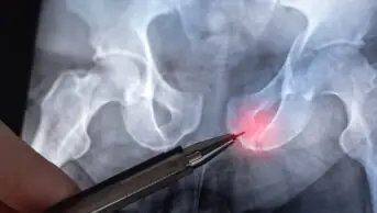
Hip fractures
Hip fractures alone account for the majority of the direct health service cost of osteoporosis. Around 50% of patients suffering a hip fracture can no longer live independently and 20% die within 12 months of the fracture[7]. Hip fractures are typically the consequence of a fall but can also occur spontaneously. For this reason, falls-related risk factors add significantly to the risk of fracture and often overlap with risk factors for osteoporosis[1].
Assessing the risk of a fragility fracture
The assessment of fracture risk is crucial in determining which patients to treat. Bone mineral density (BMD) combined with the assessment of clinical risk factors for fracture is a better predictor than BMD or clinical risk factors alone. There are a number of risk assessment tools which can predict the fracture risk over a period of time and clinicians are recommended to use these to aid decision making. There are several well established risk factors for osteoporosis including a number of drugs (See Box 1). Many of these impact on BMD but also contribute independently to future fracture risk. There are two tools which are approved by NICE for use in the UK; FRAX and QFracture. Both tools calculate the 10 year probability of major osteoporotic fracture (hip, wrist, vertebra or proximal humerus) and the 10 year probability of hip fracture.
FRAX and QFracture
The FRAX algorithm combines the patient’s age, sex, height and weight along with seven clinical risk factors (previous fracture, parent who has had a hip fracture, current smoking, glucocorticoid use, rheumatoid arthritis, secondary osteoporosis and alcohol consumption of ≥3 units per day)[8]. FRAX assessment can be performed in patients in whom BMD has not been measured, but adding a BMD result to the FRAX input data further improvesthe accuracy of the fracture prediction.
The QFracture algorithm can be used for individuals aged between 30 and 99 years and incorporates a series of risk factors. Examples of those that differ to FRAX include ethnicity, history of falls, nursing or care home residence and multiple secondary causes of osteoporosis[9]. A full list of risk factors can be accessed via the website listed in the references. In contrast to FRAX, the QFracture algorithm cannot incorporate BMD values and thresholds for treatment are less well defined. These tools, whilst being simple to use and very powerful, do have the limitation that they may not include every risk factor, or use a yes/no approach, rather than a dose dependent approach (e.g. smoking and alcohol). Clinicians must recognise that this may lead to overestimation or underestimation of actual fracture risk in some clinical circumstances.
Targeting risk assessment
The risk of fracture should be assessed:
- In all women aged 65 years and over and all men aged 75 years and over;
- In women aged under 65 years and men aged under 75 years in the presence of risk factors, for example:
- Previous fragility fracture
- Current use or frequent use of oral or systemic glucocorticoids
- History of falls
- Family history of hip fracture
- Other causes of secondary osteoporosis
- Low BMI (<18.5kg/m2)
- Smoking
- Alcohol intake of more than 14 units per week for women and more than 21 units per week for men[1].
Fracture risk should not be routinely assessed in people aged less than 50 years unless they have major risk factors (e.g. current or frequent use of glucocorticoids) because they are unlikely to be at high risk[1].
Diagnosis
The predictive value of BMD for hip fracture is at least as that of blood pressure for stroke[1]. BMD testing allows a physician to diagnose osteoporosis in high risk patients before a fracture occurs and offer interventions to reduce fracture risk. In women who have had a fragility fracture and are aged 75 years or older, osteoporosis should be assumed without the need for BMD measurement and treatment should be started immediately. Similarly men or women over the age of 64 years who have been taking oral corticosteroids for three months or more should also be treated immediately without further testing of BMD. The National Osteoporosis Guideline Group (NOGG) recommends BMD measurement in patients who are above the lower assessment threshold but below the higher assessment threshold on the FRAX prediction chart.
The International Osteoporosis Foundation and WHO recommends that the reference technology for the diagnosis of osteoporosis is dual energy X-ray absorptiometry (DXA). DXA is a technique that uses extremely low doses of ionising radiation to quantify BMD as a T-score accurately and precisely. It is also the most clinically useful way to monitor the effects of therapy. Table 2 demonstrates the BMD scale used for the diagnosis of osteoporosis. Generally BMD is performed at two sites; hip and lumbar spine (forearm is substituted if previous bilateral hip replacements or previous spinal surgery). Other sites e.g. calcaneum and validated technologies (quantitative ultrasound) may be used in clinical practice, but it should be recognised that axial BMD measurement by a standard DXA scan is the gold standard.
Table 2. Bone mineral density scale
| Category | Description | T-score |
| Normal | A value of BMD within 1 standard deviation of the young adult reference mean | ≥ -1 |
| Low bone mass (osteopenia) | A value of BMD more than 1 standard deviation below the young adult reference mean but less than 2.5 standard deviations below this mean | < -1 and >-2.5 |
| Osteoporosis | A value of BMD 2.5 standard deviations or more below the young adult reference mean | ≤ -2.5 |
| Severe osteoporosis | A value of BMD 4 standard deviations or more below the young adult reference mean | ≤ -4 |
Summary
Osteoporosis is a consequence of an imbalance in the process of bone remodelling. This results in a significant increase in the risk of fragility fractures which are associated with high mortality and morbidity as well an increasing financial burden on the National Health Service. Risk assessment is pivotal to identify individuals who are at high risk of fractures and in whom preventative treatment will provide benefit.
References
- National Institute for Health and Clinical Excellence. Osteoporosis: assessing the risk of fragility fracture. August 2012.
- National Osteoporosis guideline Group, Osteoporosis: Clinical guideline for prevention and treatment. March 2014. www.shef.ac.uk/NOGG/NOGG_Executive_Summary.pdf (Accessed 28 March 2014)
- Seeman E and Delmas PD. Bone quality –The material and structural basis of bone strength and fragility. New England Journal of Medicine 2006;354:2250-614.
- Manolagas SC. Pathogenesis of osteoporosis. In Uptodate, Post TW (Ed), Up to date, Waltham MA; 2014 (Accessed 18 November 2013)
- Whyte MP. The long and short of bone therapy. New England Journal of Medicine 2006; 354:860-8636.
- Copeland C, Worsley A. Osteoporosis features of disease and diagnosis. Pharm J 2009;1:211-2157.
- The National Hip Fracture Database. 2014. www.nhfd.co.uk (Accessed 21 March 2014)
- WHO Fracture risk assessment tool. March 2014. www.shef.ac.uk/FRAX/tool.aspx?country=1 (Accessed 29 March 2014)
- QFracture 2013 Risk calculator. March 2014. www.qfracture.org/index.php (Accessed 29 March 2014)
- Manolagas SC. Normal skeletal development and regulation of bone formation and resorption. In Up to date, Post TW (Ed), Up to date, Waltham MA;2014 (Accessed 17 March 2014).
- Manolagas SC and Jilka RL. Bone marrow, cytokines and bone remodelling. New England Journal of Medicine 1995;332:305-311.
- Lweicki ME. Managing osteoporosis: challenges and strategies. Cleveland Clinic Journal of Medicine 2009;76:457-466.