Shutterstock.com
After reading this article, you should be able to:
- Outline the principles of fracture healing;
- Describe the pharmacological management of fractures;
- Discuss measures that can be implemented to reduce the risk of future fractures.
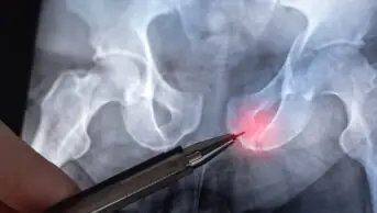
A fracture is a break in continuity of bone that occurs when force is applied to the bone that exceeds the strength of the bone itself[1]. Fracture-inducing forces may be from a single impact, such as a fall, or from repetitive loading on the bone, such as that produced by repetitive movement when running. The mechanism of injury is categorised as high-energy or low-energy impact. This is an important consideration, as it influences management choices. As well as the magnitude of the force applied to the bone, the mechanism of injury and quality of bone structure influence the risk of fracture[2]. Fracture incidence in the UK has been reported as 73.3 per 10,000 person years of follow up (all ages), although it is 116.3 per 10,000 person years of follow up when considering incidence rates in those aged 50 years and over[3].
There are approximately 47.8 fractures per 10,000 population that require hospital admission[4]. Fractures that occur following a low impact and as a result of underlying pathology are sometimes referred to as pathologic fractures[1]. Pathologic fractures occur through areas of weakened bone attributed to either primary malignant lesions, benign lesions, metastasis or underlying metabolic abnormalities, with the common factor being altered skeletal biomechanics secondary to pathologic bone.
Glucocorticoids, aromatase inhibitors, thiazolidinediones and androgen deprivation are associated with an increased risk of fracture and therefore require a proactive approach to assessing fracture risk and taking precautions to counter these effects[5].
Fractures can cause significant morbidity and mortality and are associated with immediate, early and late complications, which can lead to impaired function and affect quality of life (see Box). Pharmacy has an essential role in counselling on bone health, advising on medicines management to reduce the risk of sustaining both initial and subsequent fractures and to promote recovery when fracture does occur.
Box: Immediate, early and late complications of fracture
- Immediate complications: soft tissue damage, nerve injury, haemorrhage;
- Early complications: infection, fat embolism, pulmonary embolism, venous thromboembolism, compartment syndrome;
- Late complications: delayed union, malunion, non-union, avascular necrosis, chronic pain, complex regional pain syndrome.
Fractures can be the first symptom of diseases such as osteoporosis, Paget’s disease of bone and metastatic bone disease. Changes to the bone remodelling process can result in reduced structural integrity of the bone; for example, in osteoporosis, reduced bone mineral density and biomechanical strength of the trabeculae diminishes the structural ability of the bone to resist mechanical forces, resulting in an increased susceptibility to fracture. Thinning and increased porosity of the cortical bone further compromises the bone’s integrity and strength, exacerbating the risk of fractures.
A fracture that occurs from force equivalent to a fall from standing height or less is classed as a fragility fracture[6]. The most common sites for fragility fractures are the hip, distal radius and vertebrae[5]. There are approximately 549,000 new fragility fractures each year in the UK; however, this statistic does not include the many vertebral fractures that go undiagnosed and unreported[7]. A patient may not be aware that they have a fracture and therefore not seek diagnostic imaging, particularly if the pain is mild. Although osteoporosis significantly increases the risk of fracture, approximately two-thirds of non-vertebral fractures occur in people who do not meet the diagnostic criteria of osteoporosis (as identified by a bone mineral density dual energy X-ray absorptiometry (DEXA) scan T-score of >–2.5)[8].
The overall aim of fracture management is to achieve healing without deformity or complication, so that usual function can be resumed. Fracture management approaches seek to restore normal alignment of the bone where possible. The alignment is maintained through immobilisation techniques, such as casts, braces or surgical fixation, which allow healing and rehabilitation to restore normal function. Where underlying pathology has predisposed the patient to fracture, this should also be addressed to reduce the risk of further fracture.
Fracture classification
Fractures are typically described by stating the bone affected, anatomic location of the injury, type of fracture and if any displacement of the fracture segment has occurred. Common descriptors also include if the fracture extends across the full thickness of the bone, the stability of the injury, or if the skin was pierced by the bone at the time of injury. Common characteristics are outlined in the table below[9].
Fracture classification systems describe important clinical features that influence the course of treatment[10,11]. Fracture classifications can be used for prognostic purposes; for example, predicting the occurrence of avascular necrosis, which requires proactive management with arthroplasty.
Many classification systems are available, such as the universal AO system, the injury-specific Neer classification of displaced proximal humeral fractures, and the Garden classification of femoral neck fractures[12–14]. Although the universal system can be used to describe fractures, it is more common to use the anatomical classification systems. National Institute for Health and Care Excellence (NICE) guidance has been developed on the management of fractures (simple and complex), major trauma and spinal injury[15–18]. Simple fractures are defined as those that may be treated in the emergency department or orthopaedic clinic. Complex fractures refer to pelvic fractures, open fractures and severe ankle fractures.
Fracture healing
Fractures can heal through direct (primary) or indirect (secondary) healing processes. Primary healing is where the fracture is reduced with surgical fixation, placing the bone fragments in direct apposition and immobilising them with implants, such as plates and screws, to create a rigid fixation. This allows direct bone remodelling to occur across the fracture site without callus formation[19]. Most fractures heal by secondary healing, which takes place in four phases: inflammation, soft callus formation, hard callus formation and remodelling[19].
Inflammatory phase
Immediately following the injury, the initial proinflammatory response produces tumour necrosis factor-α (TNF-α), interleukin-1 (IL-1), IL-6, IL-11 and IL-18. These factors recruit inflammatory cells to the injury site and promote angiogenesis[20]. A haematoma is formed at the fracture site to stem further bleeding and macrophages, and mesenchymal cells migrate towards the injury. The inflammatory response can last up to 7 days, although it typically peaks at 24 hours[19].
Soft callus formation
During this phase, the area progresses from the initial inflammatory response towards establishing stability at the site. Gradually, early granulation tissue is formed, replacing the haematoma, and bone debris and necrotic tissue is reabsorbed[21,22]. During the soft callus formation stage, a fibrocartilage callus is formed, bridging the gap between the fracture sites.
Hard callus formation
At this stage, the soft fibrocartilaginous structure is hardened by the deposit of minerals, such as calcium and phosphate, initiating the transition to a rigid, hard structure. The hardening callus temporarily stabilises the fracture, allowing limited mobility while the bone continues to heal.
Remodelling
Once the fracture has unified, the bone will be gradually remodelled by osteoclasts resorbing bone and osteoblasts forming new bone, restoring normal cortical structure and removing excess periosteal callus[21]. It may take several years before the remodelling phase is complete[19].
Fracture healing considerations
Physiotherapy and occupational therapy input may be required to assist with rehabilitation of the injury and restoration of function, especially if the injury was complex or extensive. Rehabilitation and functional progress can be monitored using patient-reported outcome measures to ensure a patient-centred approach to rehabilitation is adopted.
Fracture healing may be inhibited by poor blood supply; mechanical factors, such as the location of the injury; and the extent of damage[21,23]. Patient factors that may impede healing include older age, nutritional deficiencies, infection, smoking, diabetes mellitus and prescription medication that alters bone remodelling and mineralisation processes[21,24]. Steroids, non-steroidal anti-inflammatory drugs (NSAIDs) and immunosuppressants are known to disturb the inflammatory process, impeding fracture repair[25–27]. Some concerns have been noted around anticoagulant medication having a detrimental effect on bone healing through the disruption of blood clot formation, bone remodelling and the vasculature[28,29]. Systematic analysis has identified that the use of low-molecular-weight heparin (LMWH) for three to six months did not increase the risk of fractures compared with unfractionated heparin[30]. However, since this review, additional reports have suggested that LMWH has a less detrimental effect than heparin or warfarin[28,29]. There have been concerns that bisphosphonates may result in a longer healing time or increase the risk of non-union; however, recent reviews suggest bisphosphonates are safe to initiate in the acute phase of care[27,31–33].
Low-intensity pulsed ultrasound (ultrasound therapy delivered at a lower intensity) has been found to promote fracture healing through the nanomotion (small movements) of the fracture site, leading to enhanced mineralisation and stability of the fracture site[27]. While this treatment is not routinely used for simple fractures, it may be used to promote healing where there is delayed or non-union of fracture[34].
Initial pharmacological management
Analgesia
The aim of pain management is to reduce pain, allow mobilisation and return to normal function. Severe, untreated, acute pain may predispose the patient to developing chronic pain and increase the risk of complications associated with immobility, such as pressure ulcers, venous thromboembolism and respiratory infection[35]. It is important to enquire regularly about a patient’s experience of pain and not assume that pain relief is adequate. The effectiveness of analgesia and adverse effects of the medication, such as nausea and constipation, should also be reviewed regularly.
Pain should be systematically evaluated using an assessment scale that is appropriate for the person’s cognitive function and developmental age[16]. A commonly used tool to assess pain is the visual analogue scale, which asks patients to rate pain on a scale of 0 (no pain) to 10 (worst imaginable pain). Although this helps to identify the intensity of the pain, it is also important to enquire about pain characteristics (e.g. whether it is aching, sharp, burning) as this will identify the type of pain (e.g. neuropathic or nociceptive pain) to be addressed.
Pain relief should be matched to the extent of injury and stage of recovery[36,37]. NICE recommends the use of oral paracetamol for mild pain, with the addition of codeine where the pain is moderate[14]. Severe pain should be managed initially with intravenous paracetamol alongside intravenous morphine titrated to patient response[15]. Major trauma and complex fractures should be managed with intravenous morphine, although intranasal diamorphine or ketamine may be used where intravenous access has not yet been established[16]. Caution is needed when using opioids in older adults owing to increased sensitivity and reduced renal clearance[15,38]. Nerve block injections may be used as an adjunct to assist with pain management, particularly in hip fractures[39]. A referral to pain management specialists is recommended for patients who have pre-existing, complex pain managed with high-dose opioids, owing to the potential for opioid tolerance and opioid-induced hyperalgesia. NSAIDs should only be considered as supplemental analgesia because of their potential impact on bone healing[26]. There are also concerns around NSAID use in those with impaired renal function and in those with an increased risk of gastrointestinal bleeding[15].
Some groups of patients are at risk of under-assessment and under-treatment of pain[40]. Cognitive impairment can impact the ability to accurately determine and report pain[41]. Other challenges noted with pain management, particularly in older adults, include stoicism and fear of addiction to medication[40]. Barriers to caregivers include knowledge deficits and attitudes towards pain[40].
Patients may be discharged from the hospital while still requiring opioid analgesia. This will require ongoing review as their fracture pain and surgical pain improves. Patients should be encouraged to record when they take analgesics, as this can aid pain control[36]. When analgesia is to be reduced, a reverse analgesic ladder is recommended, with opioids being reduced before paracetamol is stopped[36]. The rationale for tapering opioid regimens should be carefully discussed with the patient and arrangements made for monitoring and support during opioid tapering[35]. It is recommended that opioid and non-opioid analgesics are prescribed separately to allow dose amendments for individual analgesics, tapering by 10% weekly or over two-week periods[35]. Patients who are still taking opioids 90 days post-surgery should trigger further assessment to review for chronic pain or possible misuse[36]. Effective pain assessment is vital to ensuring the correct type and dose of medication is prescribed. Opioid-tolerant patients may need the support of specialist pain services when decreasing medication[36].
Antibiotics
Fracture-related infection (FRI) is a major complication, which can result in limb amputation owing to the burden of infection or instability caused by lytic lesions[42]. Open fractures have a high risk of infection and require the administration of prophylactic intravenous antibiotics within the emergency department if they have not previously been administered[18]. Fractures that require surgical fixation and orthopaedic implants to stabilise the fracture will also require prophylactic use of antibiotics to reduce the risk of implant and surgical-site infection[43]. The topical use of antibiotics on surgical wounds, however, is not recommended[43].
FRI requires a multidisciplinary and multifaceted treatment approach, which may include surgical debridement and antibiotic therapy[44]. When patients have signs of sepsis, blood cultures should be obtained and parenteral antibiotic therapy commenced without delay[45]. Patients suspected of having an early or acute FRI, but who are not systemically unwell, should be reviewed by a consultant within 48 hours[45]. Empiric antibiotic therapy should not be prescribed before this review, or without diagnostic intervention, as it impedes the identification of the causative organism.
Broad-spectrum antibiotics should be started after sampling, but should be narrowed down and culture-specific once this is possible[45]. Rifampicin is commonly chosen to treat staphylococcal implant-related infections; however, it must be used as part of combination therapy because of the risk of the rapid emergence of resistant microorganisms[42]. Local delivery of antimicrobial therapy may also be required to eradicate deep infection. Research to explore new bioresorbable delivery mechanisms and enhanced pharmacokinetic release is ongoing[44].
Prevention of future fractures
Fragility fractures are a significant risk factor for sustaining future fractures[46]. While measuring bone mineral density can be helpful in determining treatment approaches, it does not assess the structural integrity of the bone or any wider risks for fracture. A fracture risk assessment should be conducted in people with clinical risk factors for a fragility fracture, so that proactive measures can be taken[5]. The QFracture tool or Fracture Risk Assessment Tool (FRAX) can be used to predict the risk of future major osteoporotic fracture[6,8].
Anti-resorptive treatment
Anti-resorptive medications are used in conditions where there is excessive bone tissue resorption, such as osteoporosis. The primary goal of anti-resorptive therapy is to slow down the activity of the osteoclast cells responsible for breaking down bone tissue. By reducing bone resorption, anti-resorptive medications help to maintain or improve bone density and reduce the risk of fractures. Bisphosphonates should be offered to individuals with a bone mineral density T-score of –2.5 or lower[6]. The National Osteoporosis Guideline Group recommends oral alendronate or risedronate, or intravenous zoledronate, as the most cost-effective interventions[5]. Owing to the poor bioavailability of oral bisphosphates (0.3–1.0% of the dose absorbed), it is important to advise patients on fasting requirements and actions that can be taken to ensure optimal use[47]. Examples include taking the medication on an empty stomach, with water, and avoiding taking food or other medication for at least 30 minutes[48]. When bisphosphonates are contraindicated or poorly tolerated, alternative options may be considered, such as denosumab, raloxifene or strontium ranelate[5,6].
Anabolic treatment
Anabolic treatment refers to a therapeutic approach that aims to promote bone formation and increase bone mass. Anabolic agents are medications that stimulate the activity of the bone-forming osteoblasts, leading to the formation of new bone tissue. Teriparatide may be required for those at very high risk of fracture, while romosozumab may be considered for the treatment of post-menopausal women who have sustained a major osteoporotic fracture within the previous 24 months[5,49]. Non-osteoporotic causes of fragility fractures (e.g. metastatic bone disease, multiple myeloma, osteomalacia) should be managed according to the underlying pathology identified.
Calcium and vitamin D supplementation
Calcium and vitamin D are required for bone mineralisation. When reviewing dietary intake, it is important to consider any co-existing disease and drug therapy that may contribute to malabsorption or poor utilisation of nutrients (e.g. intestinal malabsorption syndromes, such as Crohn’s disease, may reduce the efficiency of absorption, or conditions that impair activation of nutrients (e.g. severe liver disease which inhibits production of 25-Hydroxyvitamin D), or affect the bone mineralisation process itself[6,8]. When dietary intake is inadequate, oral supplementation should be offered as an adjunct to osteoporosis treatment, particularly for those who are at risk of vitamin D deficiency[8]. If calcium intake is adequate (700mg/day), 10 micrograms (400 international units [IU]) of vitamin D can be prescribed for those who have limited exposure to sunlight. For individuals whose dietary calcium intake is inadequate, NICE recommends 20 micrograms (800 IU) of vitamin D and at least 1g of calcium daily for older or housebound patients, such as those in nursing or residential care[6]. Vitamin A is also needed for effective osteoblast function and vitamin C for collagen synthesis; these should be considered as part of a holistic nutritional assessment[50].
Supporting patients following hospital discharge
Pharmacy teams and prescribers play an essential role in fracture prevention and fracture management. They are ideally placed to offer education on modifiable risk factors for bone health (e.g. diet, exercise, smoking cessation) and can provide an ongoing holistic review of medication following discharge from hospital[8]. Reviewing adherence to medication after discharge from hospital and the effectiveness of treatment often takes place within community care. Tapering and deprescribing of medicines no longer required will also be managed by primary care following discharge. Pharmacy professionals have a vital role in supporting patients through bisphosphonate counselling and ensuring the safety and efficacy of treatment. They are also well placed to note potential adverse effects of medication on bone health and advise on actions that can be taken to promote bone health.
Falls
Falls are a common cause of fractures and require a multifactorial and multidisciplinary approach to reducing their occurrence[51]. A medication review is essential in preventing falls, particularly through the review of polypharmacy, use of psychoactive drugs and medications that can cause postural hypotension[52]. The use of validated screening tools targeted at fall prevention is recommended[51,53]. A practical approach for effective review of medication in patients at risk of falls is available from the Royal Pharmaceutical Society[54]. Alongside medication reviews, pharmacy teams can recommend activities that reduce the likelihood of falls, such as regular balance and resistance exercises. They can also play an important role in fragility-fracture risk reduction by encouraging patients to adopt healthier lifestyles, with particular reference to a balanced diet and reducing smoking and alcohol intake.
Summary
Fractures can cause significant morbidity and mortality and require a multidisciplinary approach to ensure optimal patient outcomes. While initial fracture management focuses on pain management and rehabilitation following injury, it is important to reduce the risk of future fractures. Preventative strategies include dietary and lifestyle advice on bone health, the use of anti-resorptive agents and reducing fall risks where possible.
Best practice points
- The pain of fracture patients should be assessed regularly using a structured assessment process. Analgesic choice should reflect the extent of injury and stage of recovery, and should be titrated using the analgesic pain ladder;
- Patients suspected of having a fracture-related infection should be reviewed by the orthopaedic consultant. Antibiotic therapy should not be prescribed before this review or without diagnostic intervention. Patients suspected of having sepsis should be treated immediately in accordance with sepsis protocols;
- Patients who have sustained fractures through low-impact mechanisms should be followed up with a bone health assessment and treatment of low bone mineral density as applicable. This should be combined with a fall risk assessment and medication review to proactively reduce the risk of falls and subsequent fractures;
- A fracture risk assessment should be undertaken for all patients at risk of fragility fractures, even if they have not suffered a fracture in the past.
- 1Stillwagon MR, Ostrum RF. Principles of Musculoskeletal Fracture Care. Clinical Foundations of Musculoskeletal Medicine. 2021;239–53. https://doi.org/10.1007/978-3-030-42894-5_19
- 2Komisar V, Robinovitch SN. The Role of Fall Biomechanics in the Cause and Prevention of Bone Fractures in Older Adults. Curr Osteoporos Rep. 2021;19:381–90. https://doi.org/10.1007/s11914-021-00685-9
- 3Curtis EM, van der Velde R, Moon RJ, et al. Epidemiology of fractures in the United Kingdom 1988–2012: Variation with age, sex, geography, ethnicity and socioeconomic status. Bone. 2016;87:19–26. https://doi.org/10.1016/j.bone.2016.03.006
- 4Jennison T, Brinsden M. Fracture admission trends in England over a ten-year period. annals. 2019;101:208–14. https://doi.org/10.1308/rcsann.2019.0002
- 5Clinical guideline for the prevention and treatment of osteoporosis. National Osteoporosis Guideline Group. 2021. https://www.nogg.org.uk/full-guideline (accessed November 2023)
- 6Osteoporosis – prevention of fragility fractures. National Institute for Health and Care Excellenc. 2023. https://cks.nice.org.uk/topics/osteoporosis-prevention-of-fragility-fractures/ (accessed November 2023)
- 7Harvey NC, Poole KE, Ralston SH, et al. Towards a cure for osteoporosis: the UK Royal Osteoporosis Society (ROS) Osteoporosis Research Roadmap. Arch Osteoporos. 2022;17. https://doi.org/10.1007/s11657-021-01049-7
- 8Management of osteoporosis and the prevention of fragility fractures. Scottish Intercollegiate Guidelines Network. Management of Osteoporosis and the Prevention of Fragility Fractures. 2021. https://www.sign.ac.uk/our-guidelines/management-of-osteoporosis-and-the-prevention-of-fragility-fractures/ (accessed November 2023)
- 9Walker J. Assessment and management of fractures. Br J Community Nurs. 2023;28:352–8.
- 10Audig?? L, Bhandari M, Hanson B, et al. A Concept for the Validation of Fracture Classifications. Journal of Orthopaedic Trauma. 2005;19:404–9. https://doi.org/10.1097/01.bot.0000155310.04886.37
- 11Meinberg E, Agel J, Roberts C, et al. Fracture and Dislocation Classification Compendium—2018. Journal of Orthopaedic Trauma. 2018;32:S1–10. https://doi.org/10.1097/bot.0000000000001063
- 12Neer C. Displaced proximal humeral fractures. I. Classification and evaluation. J Bone Joint Surg Am. 1970;52:1077–89.
- 13Garden RS. LOW-ANGLE FIXATION IN FRACTURES OF THE FEMORAL NECK. The Journal of Bone and Joint Surgery. British volume. 1961;43-B:647–63. https://doi.org/10.1302/0301-620x.43b4.647
- 14Fractures (non-complex): assessment and management. National Institute of Health and Care Excellence. 2016. https://www.nice.org.uk/guidance/ng38 (accessed November 2023)
- 15Major trauma: assessment and initial management. National Institute of Health and Care Excellence. 2016. nice.org.uk/guidance/ng39 (accessed November 2023)
- 16Spinal injury: assessment and initial management. National Institute of Health and Care Excellence. 2016. https://www.nice.org.uk/guidance/ng41 (accessed November 2023)
- 17Fractures (complex): assessment and management. National Institute of Health and Care Excellence. 2022. https://www.nice.org.uk/guidance/ng37/evidence/a-negative-pressure-wound-therapy-for-temporary-closure-of-open-fractures-pdf-11261965214 (accessed November 2023)
- 18Marsell R, Einhorn TA. The biology of fracture healing. Injury. 2011;42:551–5. https://doi.org/10.1016/j.injury.2011.03.031
- 19Gerstenfeld LC, Cullinane DM, Barnes GL, et al. Fracture healing as a post-natal developmental process: Molecular, spatial, and temporal aspects of its regulation. J. Cell. Biochem. 2003;88:873–84. https://doi.org/10.1002/jcb.10435
- 20Mirhadi S, Ashwood N, Karagkevrekis B. Factors influencing fracture healing. Trauma. 2013;15:140–55. https://doi.org/10.1177/1460408613486571
- 21Craig J, Clarke S, Moore P, et al. Principles of fracture management. In: Clarke S, Drozd M, eds. Orthopaedic Trauma Nursing: An Evidence-Based Approach to Musculoskeletal Care . Wiley-Blackwell 2023:221–235.
- 22Keramaris NC, Calori GM, Nikolaou VS, et al. Fracture vascularity and bone healing: A systematic review of the role of VEGF. Injury. 2008;39:S45–57. https://doi.org/10.1016/s0020-1383(08)70015-9
- 23Jiao H, Xiao E, Graves DT. Diabetes and Its Effect on Bone and Fracture Healing. Curr Osteoporos Rep. 2015;13:327–35. https://doi.org/10.1007/s11914-015-0286-8
- 24Kovach TK, Dighe AS, Lobo PI, et al. Interactions between MSCs and Immune Cells: Implications for Bone Healing. Journal of Immunology Research. 2015;2015:1–17. https://doi.org/10.1155/2015/752510
- 25Wheatley BM, Nappo KE, Christensen DL, et al. Effect of NSAIDs on Bone Healing Rates: A Meta-analysis. J Am Acad Orthop Surg. 2019;27:e330–6. https://doi.org/10.5435/jaaos-d-17-00727
- 26Foulke BA, Kendal AR, Murray DW, et al. Fracture healing in the elderly: A review. Maturitas. 2016;92:49–55. https://doi.org/10.1016/j.maturitas.2016.07.014
- 27Li Y, Liu L, Li S, et al. Impaired bone healing by enoxaparin via inhibiting the differentiation of bone marrow mesenchymal stem cells towards osteoblasts. J Bone Miner Metab. 2021;40:9–19. https://doi.org/10.1007/s00774-021-01268-5
- 28Butler AJ, Eismont FJ. Effects of Anticoagulant Medication on Bone-Healing. JBJS Reviews. 2021;9:e20.00194. https://doi.org/10.2106/jbjs.rvw.20.00194
- 29Gajic-Veljanoski O, Phua CW, Shah PS, et al. Effects of Long-Term Low-Molecular-Weight Heparin on Fractures and Bone Density in Non-Pregnant Adults: A Systematic Review With Meta-Analysis. J GEN INTERN MED. 2016;31:947–57. https://doi.org/10.1007/s11606-016-3603-8
- 30Molvik H, Khan W. Bisphosphonates and their influence on fracture healing: a systematic review. Osteoporos Int. 2015;26:1251–60. https://doi.org/10.1007/s00198-014-3007-8
- 31Palui R, Durgia H, Sahoo J, et al. Timing of osteoporosis therapies following fracture: the current status. Therapeutic Advances in Endocrinology. 2022;13:204201882211129. https://doi.org/10.1177/20420188221112904
- 32Tong YYF, Holmes S, Sefton A. Early bisphosphonate therapy post proximal femoral fracture fixation does not impact fracture healing: a systematic review and meta‐analysis. ANZ Journal of Surgery. 2022;92:2840–8. https://doi.org/10.1111/ans.17792
- 33Harrison A, Lin S, Pounder N, et al. Mode & mechanism of low intensity pulsed ultrasound (LIPUS) in fracture repair. Ultrasonics. 2016;70:45–52. https://doi.org/10.1016/j.ultras.2016.03.016
- 34Low-intensity pulsed ultrasound to promote healing of delayed-union and non-union fractures. National Institute for Health and Care Excellence. 2018. https://www.nice.org.uk/guidance/ipg623 (accessed November 2023)
- 35Opioids Aware. Faculty of Pain Medicine. 2022. https://www.fpm.ac.uk/opioids-aware (accessed November 2023)
- 36Surgery and Opioids: Best Practice Guidelines 2021. Faculty of Pain Medicine. 2021. https://fpm.ac.uk/sites/fpm/files/documents/2021-03/surgery-and-opioids-2021_2.pdf (accessed November 2023)
- 37Patient experience in adult NHS services: improving the experience of care for people using adult NHS services . National Institute for Health and Care Excellence. 2021. https://www.nice.org.uk/guidance/cg138/resources/patient-experience-in-adult-nhs-services-improving-the-experience-of-care-for-people-using-adult-nhs-services-pdf-35109517087429 (accessed November 2023)
- 38Prescribing in the elderly. British National Formulary. 2023. https://bnf.nice.org.uk/medicines-guidance/prescribing-in-the-elderly/ (accessed November 2023)
- 39Hip fracture: management. National Institute for Health and Care Excellence. 2023. https://www.nice.org.uk/guidance/cg124/resources/hip-fracture-management-pdf-35109449902789 (accessed November 2023)
- 40Jonsdottir T, Gunnarsson EC. Understanding Nurses’ Knowledge and Attitudes Toward Pain Assessment in Dementia: A Literature Review. Pain Management Nursing. 2021;22:281–92. https://doi.org/10.1016/j.pmn.2020.11.002
- 41Wennberg P, Möller M, Sarenmalm EK, et al. Evaluation of the intensity and management of pain before arrival in hospital among patients with suspected hip fractures. International Emergency Nursing. 2020;49:100825. https://doi.org/10.1016/j.ienj.2019.100825
- 42Depypere M, Morgenstern M, Kuehl R, et al. Pathogenesis and management of fracture-related infection. Clinical Microbiology and Infection. 2020;26:572–8. https://doi.org/10.1016/j.cmi.2019.08.006
- 43Surgical site infections: prevention and treatment. National Institute for Health and Care Excellence. 2020. https://www.nice.org.uk/guidance/ng125 (accessed November 2023)
- 44Foster AL, Moriarty TF, Trampuz A, et al. Fracture-related infection: current methods for prevention and treatment. Expert Review of Anti-infective Therapy. 2020;18:307–21. https://doi.org/10.1080/14787210.2020.1729740
- 45Fracture Related Infections. British Orthopaedic Association. 2019. https://www.boa.ac.uk/static/dee7cba7-5919-4f26-a286033fcf46a458/boast-fracture-related-infections.pdf (accessed November 2023)
- 46Kanis JA, Johnell O, De Laet C, et al. A meta-analysis of previous fracture and subsequent fracture risk. Bone. 2004;35:375–82. https://doi.org/10.1016/j.bone.2004.03.024
- 47Inderjeeth C, Inderjeeth A, Raymond W. Medication selection and patient compliance in the clinical management of osteoporosis. Aust Fam Physician. 2016;45:814–7.
- 48Bisphosphonates. Versus Arthritis. 2023. https://www.versusarthritis.org/about-arthritis/treatments/drugs/bisphosphonates/ (accessed November 2023)
- 49Romosozumab for treating severe osteoporosis. National Institute for Health and Care Excellence. 2022. https://www.nice.org.uk/guidance/ta791 (accessed November 2023)
- 50Anatomy and Physiology in Health and Illness. Elsevier 2018.
- 51Montero-Odasso M, van der Velde N, Martin FC, et al. World guidelines for falls prevention and management for older adults: a global initiative. Age and Ageing. 2022;51. https://doi.org/10.1093/ageing/afac205
- 52Falls – risk assessment. National Institute for Health and Care Excellence. 2019. https://cks.nice.org.uk/topics/falls-risk-assessment (accessed November 2023)
- 53Seppala LJ, Petrovic M, Ryg J, et al. STOPPFall (Screening Tool of Older Persons Prescriptions in older adults with high fall risk): a Delphi study by the EuGMS Task and Finish Group on Fall-Risk-Increasing Drugs. Age and Ageing. 2020;50:1189–99. https://doi.org/10.1093/ageing/afaa249
- 54Medicines and Falls. National Falls Prevention Coordination Group. 2023. https://www.rpharms.com/Portals/0/RPS%20document%20library/Open%20access/Pharmacy%20guide%20docs/Medicines%20and%20falls%209%2023%20(RPSendorsed).pdf?ver=kHy696ZEbkwR7eopGgbkFw%3d%3d (accessed November 2023)
You must score at least 70% in this module to pass. Ensure you have read ‘Pharmaceutical considerations for fracture management in adults’ before attempting to complete the module. This module can be taken up to three times. Once you have passed, you can view the correct answers and download a certificate as a record of your learning. Additional CPD modules are available in the ‘My CPD’ section of your account.


