

PEAKSTOCK / SCIENCE PHOTO LIBRARY
After reading this article, you should be able to:
- Explain the causes, signs and symptoms of glaucoma;
- Outline current management strategies.
Glaucoma is an eye disorder in which the optic nerve is damaged where it leaves the eye. Damage to the optic nerve is irreversible and progressive damage can lead to sight loss. Glaucoma is responsible for 16% of patients registered as sight impaired (partially sighted) or severely sight impaired (blind) in the UK. There is no cure for glaucoma but there are several effective treatments available that can preserve vision and prevent vision loss when identified early.
Glaucoma is often characterised by raised intraocular pressure (IOP). The condition is usually asymptomatic; age, race, family history and diabetes are risk factors. Around 2% of those aged over 40 years in the UK have glaucoma.
The aim of treatment is to reduce IOP. Pharmacists can play an important role in the management of the condition by promoting eye tests for screening and supporting patients with ocular treatments used to control pressure in the eye to reduce the risk of sight loss. This article will give a overview of the condition and current treatments to help enable pharmacists to support patients with glaucoma.
Aetiology
Glaucoma is a range of disorders usually characterised by raised IOP and leads to damage of the optic nerve. Damage to the optic nerve can occur with a normal IOP.
Glaucoma can be classified as primary or secondary. Primary glaucoma includes primary open angle glaucoma (chronic simple glaucoma), the most common form of glaucoma, and primary angle closure glaucoma, which is caused by blockage in the trabecular meshwork. This drains the anterior chamber of the eye to the episcleral veins through the Schlemm’s canal.
Primary angle closure glaucoma — which results from decreased outflow of aqueous humour, causing accumulation of fluid in the eye — is generally acute in onset and may require treatment as a medical emergency.
Secondary glaucomas can result from a wide range of causes, such as inflammation, tumours or congenital abnormalities.
This guide will focus on the most common form of glaucoma: primary open-angle glaucoma.
Table 1 describes the types of glaucoma[1].
Epidemiology
Causes of primary open angle glaucoma are not always clear but risk factors include:
- Age: glaucoma affects 2 in 100 of people aged over 40 years, and 1 in 10 of people aged over 75 years;
- Race: Black people have greater incidence, earlier onset and greater severity;
- Family: 10% likelihood of first-degree relatives developing glaucoma;
- Short sight (myopia): people who are short-sighted (distant object appear blurred) are prone to glaucoma;
- Diabetes: believed to increase the risk of developing glaucoma;
- Cardiovascular disease and hypertension: believed to cause a small increase in risk.
In the UK, people aged over 40 years who have an immediate family member who has been diagnosed with glaucoma are entitled to free annual sight tests.
Signs and symptoms
Glaucoma is usually asymptomatic, unless it is in the advanced stages. Open angle glaucoma usually develops slowly over several years so many patients will not experience any noticeable symptoms.
Symptoms from optic nerve damage in advanced stages include worsening of vision (tunnelling of vision/defects of the visual field).
Signs can be present before optic nerve damage develops and include raised IOP and increased variation in IOP.
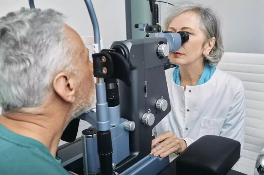
Investigations
Most patients are diagnosed at routine eye appointments:
- IOP measured by tonometry;
- Appearance of the optic nerve observed with ophthalmoscopy or a computer-based image analysis;
- Peripheral vision assessment taken using perimetry (spot testing);
- Assessment of the anterior chamber angle depth (gonioscopy and slit lamp).
It is important that all three tests are carried out to confirm diagnosis.
Management
The aim of treatment is to reduce IOP to prevent damage to the optic nerve. Treatment cannot reverse nerve damage or vision loss that has already occurred.
First line treatment for an IOP ≥ 24 mmHg is 360° selective laser trabeculoplasty (SLT). This increases drainage of the trabecular meshwork via a laser targeted at melanin rich cells. This has shown to be cost effective compared with eye drop use over time. It delays the need for eye drops but does not remove the chance of their use at a later date. Some patients may need a second SLT later if the effect on the IOP reduces over time.
Moorfields Hospital produces a patient information leaflet on SLT, which can be accessed here.
If SLT is not suitable or a patient chooses not to have SLT or further IOP lowering is required after a SLT, a generic prostaglandin analogue (e.g. latanoprost) should be used.
Other classes of pharmacological agents can be used if prostaglandin analogues are not tolerated or do not lower IOP sufficiently. The mechanism of action and possible adverse effects are seen in Table 2.
If further treatment is required after using medicines from two classes, SLT or glaucoma surgery with mitomycin C should be considered.
Monitoring parameters
Tonometry, ophthalmoscopy and perimetry should be monitored for effectiveness of treatment.
Pharmacists should monitor adherence to treatment and check eye drop installation technique.
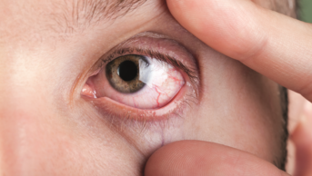
The appearance of the eye/s should also be checked for allergic reactions to treatment with eye drops[2], which include:
- Severely itchy eyes and eyelids;
- Red injected eyes;
- Red and swollen eyelids;
- Symptoms and signs that are worse after the drops are instilled, which resolve when treatment is stopped.
The frequency of reassessment should be guided by progression and clinical risk.
Although rare, systemic side effects may occur in susceptible individuals with any of the five classes of drug. Those at risk of systemic side effects can be advised to perform punctal occlusion (the compression of the tear ducts by pressing a finger to the inner corner of the eye) for two minutes after using eye drops.
Counselling
The pharmacist has an important role to play — they can help support treatment adherence, advise on eye drop technique and encourage patients to have regular eye tests and check-ups.
Patients should wash their hands before applying eye drops and the tip of the container should not touch their eyes or areas around the eye. Contamination with bacteria can cause eye infections, which have the potential to cause loss of vision.
When advising on the administration of eye drops, the following points should be covered:
- Tilt head back and pull the lower eyelid down slightly to form a pocket between the eyelid and eye;
- Invert the container and press lightly with thumb and forefinger until a single drop is instilled into the eye;
- Replace the cap immediately after use.
If more than one preparation is being used, they should be administered at least ten minutes apart.
If a patient reports issues with administering drops, observe administration to see if a drop aid device will assist the patient.
This article was adapted from FASTtrack: Therapeutics, published by Pharmaceutical Press. It was updated in March 2023 by Natalie Lewis, Education Operations Manager School of Pharmacy, Aston University.
Last updated March 2023
- 1World Glaucoma Week 2021. Metro Health HMO. 2021.https://www.metrohealthhmo.com/world-glaucoma-week-2021 (accessed Mar 2023).
- 2Glaucoma: Scenario — Primary open angle glaucoma and ocular hypertension. National Institute for Health and Care Excellence. 2022.https://cks.nice.org.uk/topics/glaucoma/management/primary-open-angle-glaucoma-intraocular-hypertension/#allergy-to-eye-drops (accessed Mar 2023).
1 comment
You must be logged in to post a comment.


Great article, brief and very useful.