SCIENCE PHOTO LIBRARY
After reading this article, you should be able to:
- Know the causes and risk factors for cellulitis, including how these risk factors can be managed and controlled;
- Recognise the symptoms of cellulitis, and know how the condition is diagnosed and any considerations for urgent referral;
- Understand the treatment options for managing cellulitis, including considerations for antibiotic prescribing and self-care options.
Introduction
Cellulitis is a common acute bacterial skin infection of the dermis and associated subcutaneous tissue, affecting around 1 in 40 people globally per year[1]. Cellulitis is characterised by pain, warmth, swelling and erythema (i.e. abnormal redness of the skin). The condition tends to respond to treatment with antibiotics, but more serious cases may require surgery to remove dead tissue and prevent infection entering the bloodstream[1]. Infections of the skin and subcutaneous tissue, including cellulitis, accounted for almost 225,000 hospital admissions in England and Wales in 2020[2]. Additionally, cellulitis and other soft tissue infections account for 18% of antibiotic prescriptions outside inpatient hospital care, second only to respiratory conditions; therefore, reducing acute infection — and thereby reducing antibiotic prescribing where possible — is an important measure in reducing antibiotic resistance[3].
Pharmacists are often in patient-facing roles and should be able to recognise the signs and symptoms of cellulitis to ensure the patient receives crucial timely treatment. This article will cover the causes and risk factors of cellulitis, as well as outlining signs and symptoms, considerations for diagnosis and the available treatment options.
Causes and risk factors
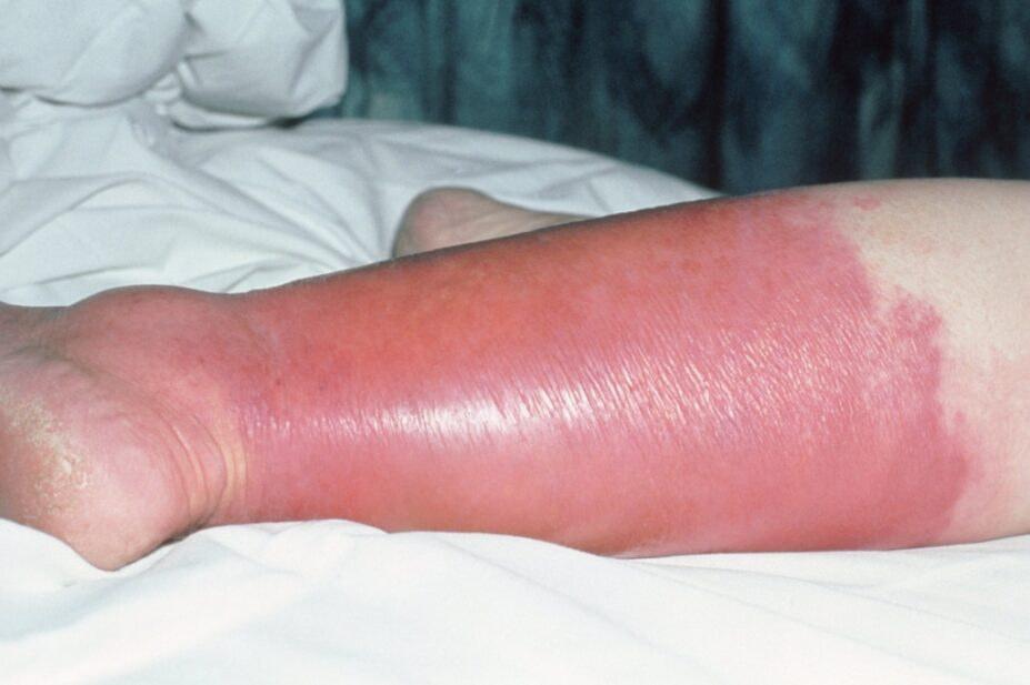
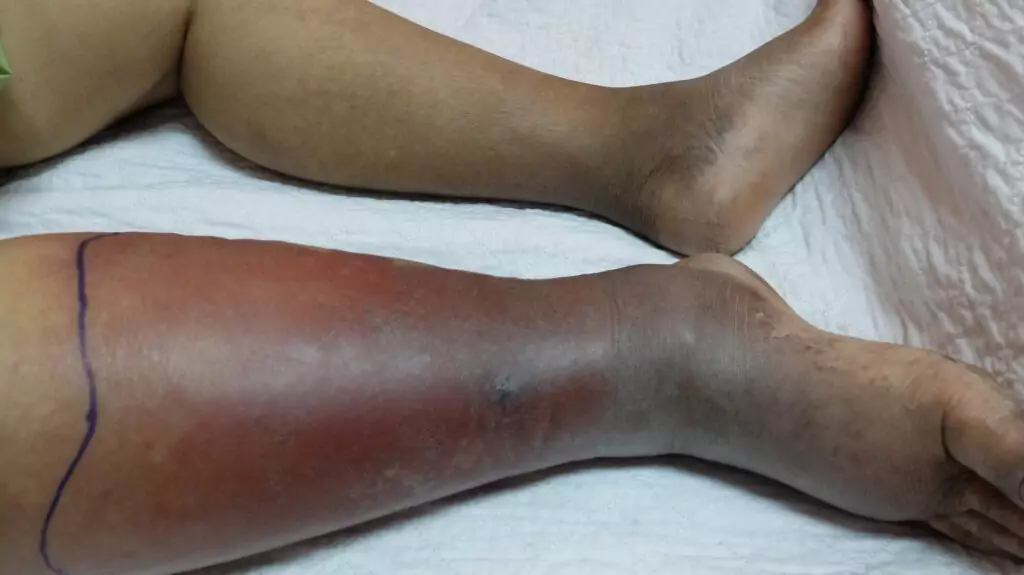
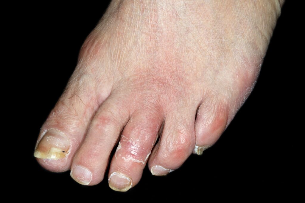
The most common pathogens involved in cellulitis are Streptococcus and Staphylococcus species[4]. The infection most commonly affects the lower limbs, reasons for which are thought to include chronic oedema, circulatory deficiency or uncontrolled eczema, the latter of which can result in dry, broken skin and entry of infection. Tinea pedis fungal infection, such as Athlete’s foot, can also cause skin breakage between the toes, leading to infection entry. Trauma to the leg, ulceration and a body mass index (BMI) of >30kg/m can cause leg swelling, leading to cellulitis; however, cellulitis can affect any limb or part of the body[5,6].
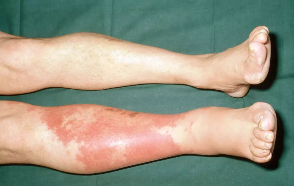
Limb infection is usually unilateral; cases of bilateral infection are rare and a differential diagnosis should be considered, such as stasis dermatitis or contact dermatitis[7]. Risk factors for cellulitis include diabetes, skin wounds, insect and animal bites, older age (individuals aged over 65 years), immune suppression, neutropenia and immunosuppressive medications[8].
Signs and symptoms
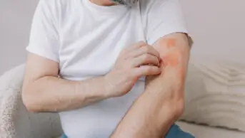
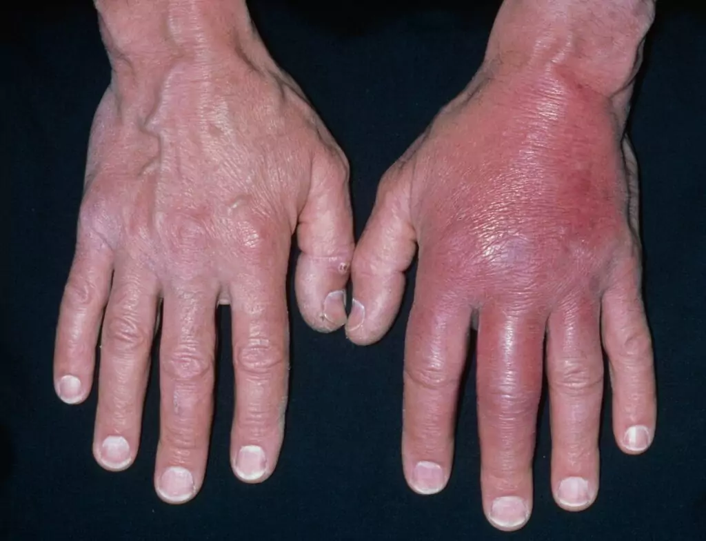
Patients may present in primary care services, such as community pharmacy or their GP, with an area affected by cellulitis, which is typically warm, swollen, tender, painful and erythematous with a poorly defined border. More severe cases can present with systemic effects such as fever, malaise, nausea, rigor and skin changes, such as blisters and bullae (i.e. large blisters that are filled with clear fluid), which would require urgent referral to hospital owing to the increased risk of sepsis[4].
Signs of cellulitis, such as erythema, are more easily noticed in lighter skin tones — more information can be found in ‘Recognising common skin conditions in people of colour’. Erythema may be subtle in darker skin tones and a faint pink, deep red, violaceous, or dark brown plaque may be seen. The presence of accompanying tenderness can help with diagnosis[9].
Shutterstock.com
Clinical Photography/SCIENCE PHOTO LIBRARY
DR P. MARAZZI/SCIENCE PHOTO LIBRARY
Cellulitis differs from erysipelas, another bacterial infection that commonly affects infants and older people with similar risk factors to cellulitis. It affects the upper dermis and superficial lymphatics. It is a superficial form of cellulitis as it involves only the upper dermis and superficial lymphatic, whereas cellulitis affects the subcutaneous tissue and dermis[7,10].
Diagnosis
Taking a thorough history will help with diagnosis and understanding how long symptoms have been present, see Box[11].
Box: Questions to aid cellulitis diagnosis
- When did the symptoms start?
- How did they start?
- How does the patient feel?
- Is the patient experiencing fevers, headaches and night sweats or general malaise?
It is also important to ascertain history of skin conditions, such as eczema or skin breach, including trauma, insect bites, fungal infections or recent surgery.
If the skin is broken and purulent, swabs should be taken[11]. Skin scrapings and nail clippings should be taken if fungal infection is suspected[11].
The initial infection shows mild redness that quickly progresses to tenderness and oedema.
The affected area for a portal of entry, such as an open wound, should be examined. When diagnosing cellulitis, the interweb spaces (between toes) should be checked for fissuring, maceration and scale; this is to ensure it is free of fungal infections, such as Athlete’s foot, and more broadly the skin should be inspected for insect bites as this can be an entry point of infection[10]. A differential diagnosis should be ruled out, such as DVT, varicose eczema, chronic inflammation secondary to lymphoedema/chronic oedema or a drug eruption[11]. The severity of cellulitis can be classified using the Eron classification system (see Table 1)[1,12].
DR P. MARAZZI/SCIENCE PHOTO LIBRARY
Class III and IV cellulitis, and cases that involve an extensive affected area that is spreading quickly, all warrant urgent hospital admission. Additionally, patients aged under one year, and individuals who are frail or immunocompromised, should be referred for urgent admission[4].
Treatment
Class I cellulitis can be treated in primary care with high-dose oral antibiotics[4]. If there is no substantial improvement after seven days of treatment with first-line oral antibiotics and the person is systemically well, adherence should be assessed and a further seven days of first-line oral treatment continued — see Table 2 for antibiotic prescribing guidance[4].
Class II cellulitis may not require admission if there are facilities and expertise available in the community setting to give intravenous antibiotics and monitor the patient — local guidelines should be reviewed[4,13]. However, management of skin infection within the community has been shown to be safe, effective and cost effective, as it reduces the need for hospital stays[14].
If the patient is deteriorating after two to three days, hospital admission should be considered, after which point further treatment will depend on local guidelines[4]. If symptoms are not improving or are worsening after 14 days, then hospital admission should be considered and specialist advice and local hospital guidelines should be followed[4].
Table 2 shows recommended antibiotic prescribing for adults aged 18 years and over with cellulitis[15].
The area of cellulitis should be marked with permanent markers by the patient to track the spread of infection. Over-the-counter analgesics can be used for pain relief — usually paracetamol or ibuprofen, depending on the patient’s medication history. Hydration is also advised and if leg(s) are involved then elevation is encouraged to promote reduction of swelling; however, compression should be avoided[4]. Additionally, the skin should be kept clean and well moisturised, antiseptic cream can be considered for cleaning cuts and wounds, and wearing appropriate clothing/footwear will help to minimise cuts and wounds[16]. The underlying risk factors of cellulitis should also be managed and controlled[4].
Risk factors for recurrent cellulitis include:
- Chronic oedema: to minimise this risk, compression bandages or stockings should be considered once the pain and infection settles;
- Dry skin/dermatitis: this can result in cracking skin or breaks in the skin, which can result in infection. It is important to keep the skin well hydrated with regular moisturising of the skin with emollients;
- Obesity: patients can be supported to reduce weight through changes in diet and exercise;
- Immunosuppression: immunosuppressed patients may require a low dose of preventative antibiotics, such as phenoxymethylpenicillin, if they experience recurrent infections[10,17].
In patients with diabetes, it is appropriate to ensure blood glucose control to minimise the risk of recurrence.
- 1Phoenix G, Das S, Joshi M. Diagnosis and management of cellulitis. BMJ. 2012;345:e4955–e4955. doi:10.1136/bmj.e4955
- 2Samannodi M. Hospital Admissions Related to Infections and Disorders of the Skin and Subcutaneous Tissue in England and Wales. Healthcare. 2022;10:2028. doi:10.3390/healthcare10102028
- 3Santer M, Lalonde A, Francis NA, et al. Management of cellulitis: current practice and research questions. Br J Gen Pract. 2018;68:595–6. doi:10.3399/bjgp18x700181
- 4Cellulitis – acute. National Institute for Health and Care Excellence. 2023.https://cks.nice.org.uk/topics/cellulitis-acute (accessed Sep 2023).
- 5Mistry K, Sutherland M, Levell NJ. Lower limb cellulitis: low diagnostic accuracy and underdiagnosis of risk factors. Clin Exp Dermatol. 2019;44:e193–5. doi:10.1111/ced.13930
- 6Bjornsdottir S, Gottfredsson M, Thorisdottir AS, et al. Risk Factors for Acute Cellulitis of the Lower Limb: A Prospective Case-Control Study. Clinical Infectious Diseases. 2005;41:1416–22. doi:10.1086/497127
- 7Brown B, Hood W. statpearls. Published Online First: 7 August 2023.http://www.ncbi.nlm.nih.gov/books/NBK549770/
- 8Njim T, Aminde LN, Agbor VN, et al. Risk factors of lower limb cellulitis in a level-two healthcare facility in Cameroon: a case-control study. BMC Infect Dis. 2017;17. doi:10.1186/s12879-017-2519-1
- 9Recognising common skin conditions in people of colour. Pharmaceutical Journal. 2021. doi:10.1211/pj.2021.1.113627
- 10Cellulitis and erysipelas patient information leaflet. British Association Of Dermatologists. 2021.https://www.bad.org.uk/pils/cellulitis-and-erysipelas/ (accessed Sep 2023).
- 11Sutherland M, Parent A. Diagnosis and management of cellulitis: a dermatology perspective. Br J Community Nurs. 2017;22:272–5. doi:10.12968/bjcn.2017.22.6.272
- 12Skin and soft tissue infections: risk factors and presentations. Pharmaceutical Journal. 2023. doi:10.1211/pj.2023.1.191226
- 13Clinical pathways: cellulitis in adults where IV antibiotic in the community may be appropriate. NHS. 2020.https://clinical-pathways.org.uk/sites/default/files/guidance/Infection/cellulitis-in-adults-where-iv-antibiotic-in-the-community-may-be-appropriate-18-may-20-v14.2.pdf (accessed Sep 2023).
- 14Nathwani D. The Management of Skin and Soft Tissue Infections: Outpatient Parenteral Antibiotic Therapy in the United Kingdom. Chemotherapy. 2000;47:17–23. doi:10.1159/000048564
- 15Cellulitis and erysipelas: antimicrobial prescribing. National Institute for Health and Care Excellence. 2019.https://www.nice.org.uk/guidance/ng141/chapter/Recommendations (accessed Sep 2023).
- 16Cellulitis. NHS. 2021.https://www.nhs.uk/conditions/cellulitis/ (accessed Sep 2023).
- 17Guidelines on the Management of Cellulitis in Lymphoedema. British Lymphology Society and the Lymphoedema Support Network. 2022.https://www.lymphoedema.org/wp-content/uploads/2022/10/management_cellulitis.pdf (accessed Sep 2023).